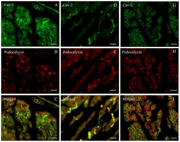
Fig. 1. Paraffin sections of mouse heart atrial tissue (wild-type).
A–I. Double label immunoflourescnce using anti caveolin-1, -2, and -3 specific antibodies (FITC-coupled secondary antibodies) and blood vessel marker anti-podocalyxin (Texas Red-coupled secondary antibody). Note the presence of Cav-1 and -3 in atrial cardiomyocytes as well as of Cav-1 and -2 in blood vessel endothelial cells. Bar = 80 μm

