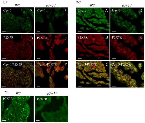
Fig. 2. Paraffin sections of mouse heart atria (A–C: WT; D–F cav-1−/−).
2/1A–F. Double label immunofluorescence for cav-1 (FITC) and P2X7R (Texas Red). The P2X7R is present in endothelial cells and in cardiomyocytes. Note the higher immunoreactivity for P2X7R in cardiomyocytes of cav-1−/− cardiac tissue. Bar = 80 μm
2/2A–F. Double label immunofluorescence for Cav-3 (FITC) and P2X7R (Texas Red). The P2X7R is present in endothelial cells and in cardiomyocytes. Note the increased expression of Cav-3 in cardiomyocytes of cav-1−/− cardiac tissue.
2/3 Control experiment demonstrates that the immnoreactivity of the anti P2X7R antibody is abolished in atrial cardiomyocytes of the corresponding knockout tissue. Bar = 80 μm

