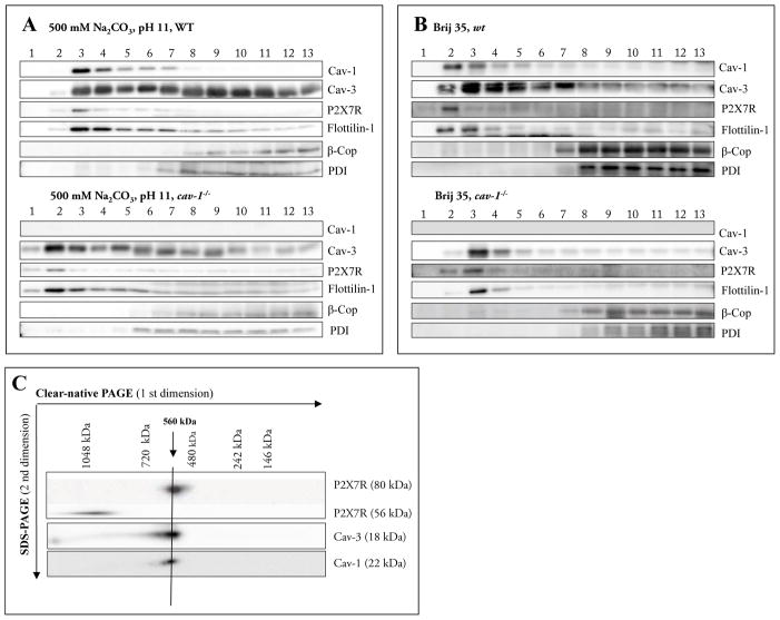
Fig. 3.
A–C Characterization of membrane fractions prepared by sonication (3A) or by Brij35 (3B) in cell homogenates of cardiac tissue from wild-type and cav-1−/− mice.
The cardiac tissue was homogenized in a buffer containing 500 mM Na2CO3 (pH11) and raft and non-raft membranes were prepared by sonication followed by centrifugation in a discontinuous sucrose gradient. Futhermore cardiac tissue was homogenized in a buffer containing 1%Brij35 and subjected to sucrose density gradient centrifugation. Thirteen fractions were collected (fraction 1, top of the gradient; fraction 13, bottom of the gradient), and an aliquot of each fraction (20 #l) was resolved by SDS-PAGE and subjected to Western blot analysis with antibodies against Cav-1, Cav-3, P2X7R, Flottilin-1, β-Cop and PDI. As expected, Cav-1, Cav-3 and flotillin-1 were enriched in fractions 3–6 (wild-type) or 1–5 (cav- 1−/−), representing caveolae-enriched membrane fractions. Representative data from three separate experiments are shown.
C Supramolecular organization of P2X7R, Cav-1 and Cav-3.
Native membrane extracts from cardiac tissue from wild-type mice were isolated and solubilized with digitonin. Proteins were separated by hrCN-PAGE/SDS-PAGE, blotted and probed with antibodies against P2X7R, Cav-1 and Cav-3. Sizes of the molecular mass standards are indicated.

