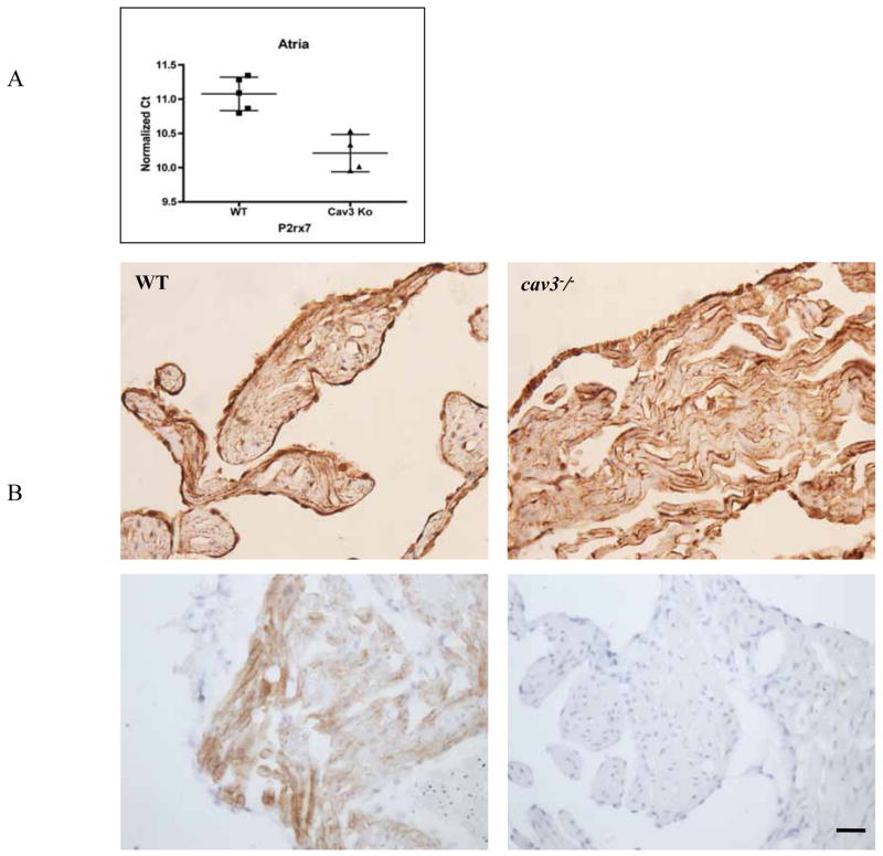
Fig. 6. Protein and mRNA measurements in cav-3−/− mice.
A P2X7R quantitative mRNA expression in atrial tissue was assessed. Cycle threshold (Ct) values were normalized to GAPDH expression. Lower Ct value equates to higher mRNA expression.
B Immunoperoxidase demonstration of P2X7R (upper panel) and Cav-3 (lower panel; immunohistochemical control experiment for the specificity of the anti-Cav-3 antibody) in atrial tissues of mouse heart (left: wild-type; right: cav-3−/− ).

