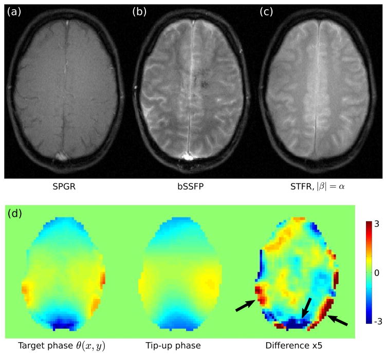
Figure 8.
Comparison of (a) SPGR (TR=10.9 ms), (b) bSSFP (TR=9.5 ms), and (c) the proposed STFR sequence (|β|= α, TR=11.1 ms). The tip-down excitation pulse was the same for all acquisitions (α = 8°). In (c), the tip-up pulse consisted of a train of 10 blips, similar to the pulse sequence shown in Fig. 1(b). SPGR (a) produces relatively low tissue signal, consistently dark CSF, and bright vessel signal due to in-flow enhancement. Balanced SSFP (b) produces bright CSF in accordance with its high T2/T1 ratio, and high vessel signal. STFR produces good gray/white matter contrast similar to bSSFP, indicating that the tip-up pulse introduces T2-weighting. (d) Comparison of target phase θ (x, y) (left), and the simulated phase of the tip-up pulse (units of radians). The phase of the tip-up pulse is in good agreement with the target phase, as desired.

