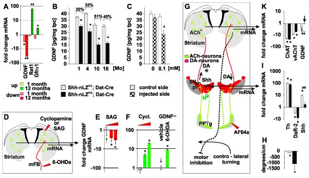
Figure 6. Shh signaling inhibits GDNF expression in the striatum.
(A) Changes in GDNF, Ret and Gfra1 gene expression in the striatum of 4 weeks and 12 months old Shh-nLZC/C/Dat-Cre mice (* p < 0.01, ** p < 0.001, two tailed t-test; n=5/genotype).
(B) Quantification of striatal GDNF tissue content as a function of age in Shh-nLZC/C/Dat-Cre mice (* p<0.01, unpaired t-test; n=10-11/group).
(C) Injection of AF64a reduces GDNF tissue content. (37 +/− 3 %; * p<0.05, unpaired t-test, n=12-14/group).
(D) Experimental design: Comparative analysis of GDNF expression upon unilateral injection of SAG or cyclopamine into the striatum of wt mice, or 6-OHDA into the mFB of GDNF-LZ+/− mice.
(E) SAG decreases GDNF mRNA expression (* p < 0.05, ANOVA followed by Tukey’s post hoc Test; n=5/group/drug concentration).
(F) Injection of cyclopamine into the striatum or 6-OHDA into the mFB increases GDNF mRNA expression (* p < 0.05, ANOVA followed by Tukey’s post hoc Test; n=5/group/drug concentration; the up-regulation caused by cyclopamine was dose dependent).
(G) Experimental design: Comparative analysis of gene expression in the vMB and striatum upon unilateral injection of AF64a into the PPTg.
(H) Quantification of turning bias 36 h post unilateral injection of AF64a into the PPTg in Shh-nLZC/C/Dat-Cre mice (* p < 0.05, post hoc test after ANOVA, n=5/genotype).
(I, K) Quantification of changes in gene expression in the vMB (I) or striatum (K) upon AF64a injection into the PPTg of Shh-nLZC/C/Dat-Cre and control mice (** p < 0.001, AF64a x genotype ANOVA followed by Tukey’s post hoc Test, (n=5/group/genotype).

