Figure 5.
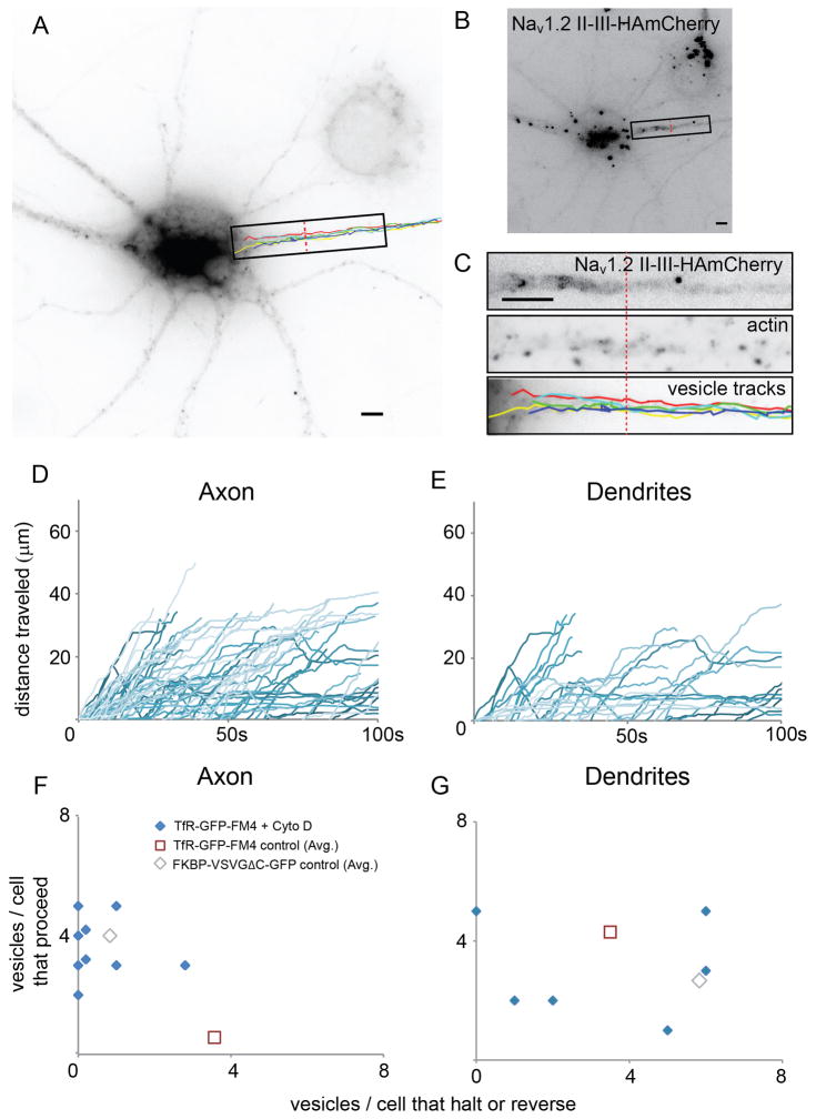
Actin filaments are necessary for halting and reversing of vesicles carrying TfR-GFP-FM4 in the AIS (see also Figure S4 and Movie 6). (A) Paths taken by vesicles carrying TfR-GFP-FM4 in the axons of cortical neurons in culture that were exposed to 4 μM cytochalasin D for 80 minutes. (B) AIS is defined by staining of Nav1.2 II-III-HAmCherry. (C) High power view of boxed region from (A) showing Nav1.2 II-III-HAmCherry, Phalloidin staining in AIS consistent with disrupted actin filaments, and vesicle tracks. (D) Plots of distance traveled versus time for vesicles in axons containing TfR-GFP-FM4 in cells exposed to cytochalasin D are consistent with those vesicles moving beyond the AIS into the distal axon. (E) Plots of distance versus time for vesicles in dendrites containing TfR-GFP-FM4 in cells exposed to cytochalasin D are similar to comparable plots of similar vesicles in control cells. (F, G) Axon and dendrite scatter plots showing the ratio of the number of vesicles carrying TfR-GFP-FM4 that proceed versus the number that halt or reverse for cells exposed to cytochalasin D. Note that each data point refers to a single cell. In axons such vesicles behave differently from similar vesicles in control cells, but similarly to those carrying FM4-VSVGΔC-GFP. In dendrites, vesicles carrying TfR-GFP-FM4 in cells exposed to cytochalasin D do not behave in a noticeably different manner from similar vesicles in control cells. Scale bar is 5 μm.

