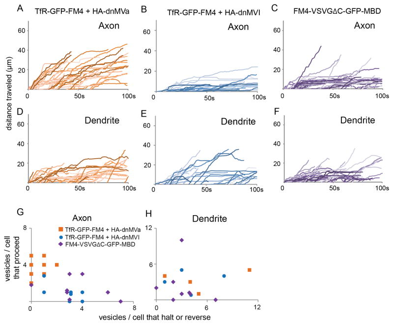
Figure 6.
Interaction with Myosin Va, but not Myosin VI, is necessary for vesicle halting and reversing, whereas interaction with Myosin Va is sufficient to cause halting of vesicles at the AIS. (see also Figure S5 and Movies 7, 8). (A) Plots of distance traveled versus time for vesicles in axons containing TfR-GFP-FM4 in cells co-expressing HA-dnMVa are consistent with those vesicles moving beyond the AIS into the distal axon. In contrast, similar plots for vesicles in the axon containing TfR-GFP-FM4 in cells coexpressing HA-dnMVI (B) indicate that these vesicles are likely to halt or reverse while in the AIS. Note that this pattern is similar to that of cells expressing TfR-GFP-FM4 only. Similarly, plots of distance traveled versus time for vesicles in axons carrying FM4-VSVGΔC-GFP-MBD (C) indicate that those vesicles are more likely to halt within the AIS than vesicles carrying FM4-VSVGΔC-GFP. Plots of distance versus time show that vesicles in dendrites containing TfR-GFP-FM4 behave similarly in cells co-expressing HA-dnMVa (D) or HA-dnMVI (E). In addition, both types of vesicles behave in a similar manner to vesicles carrying FM4-VSVGΔC-GFP-MBD (F) in dendrites of control cells. (G, H) Axon and dendrite scatter plots showing the ratio of the number of vesicles carrying TfR-GFP-FM4 that proceed versus the number that halt or reverse in cells co-expressing HA-dnMVa or HA-dnMVI and a similar ratio for vesicles carrying FM4-VSVGΔC-GFP-MBD in control cells. Note that each data point refers to a single cell. In axons the vesicles carrying TfR-GFP-FM4 in cells co-expressing HA-dnMVI and vesicles carrying FM4-VSVGDC-GFP-MBD in control cells behave similarly to each other, but differently from those carrying TfR-GFP-FM4 in cells co-expressing HA-dnMVa. All three types of vesicles behave similarly in the dendrites.

