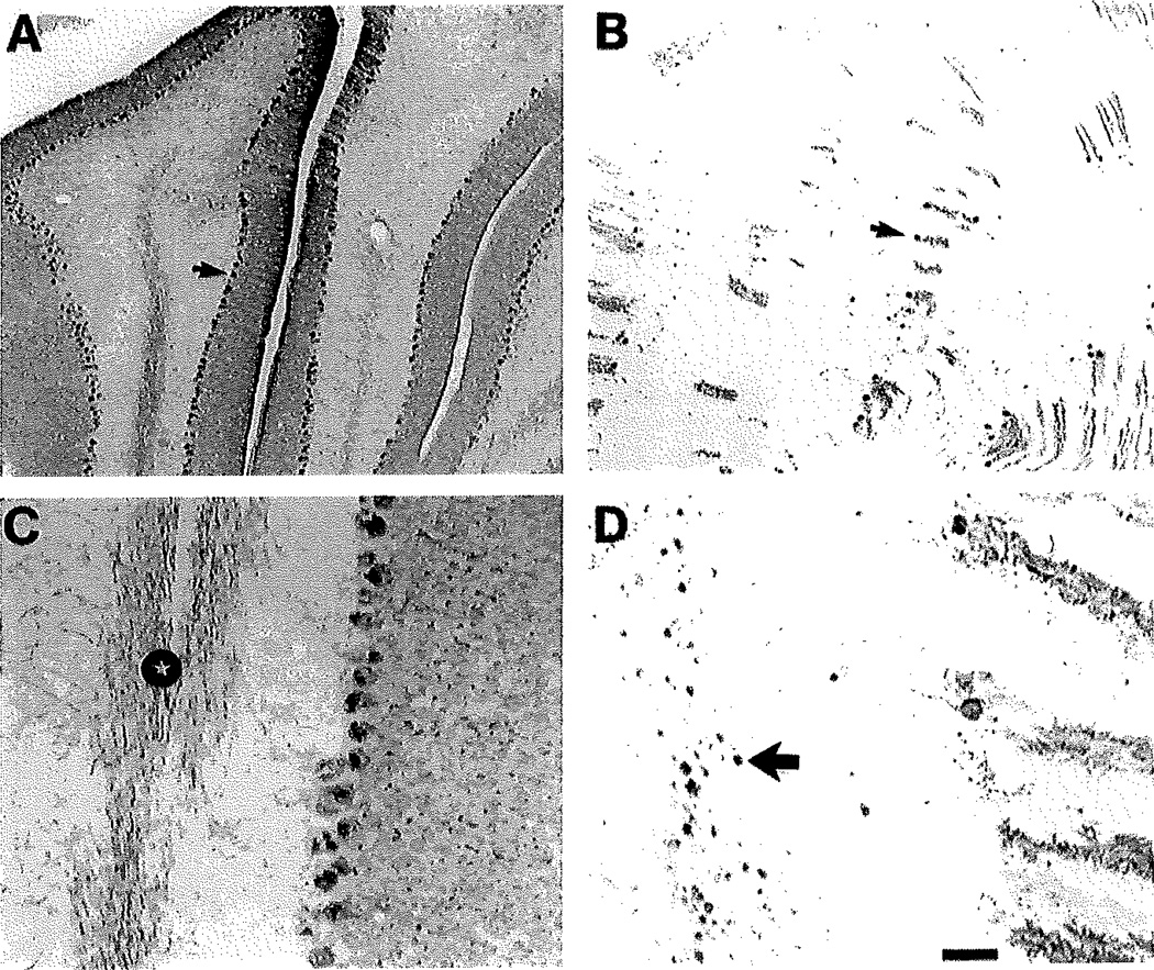
Fig. 2.
Calbindin-immunostained sections illustrating the loss of cerebellar Purkinje cells in the 11-week-old NPC−/− mouse. Calbindin is a selective marker for Purkinje cells in the cerebellum, their dendrites and axons. A: In the 11-week-old NPC+/+ mouse there are numerous Purkinje cell closely spaced together (arrow points to the cell body of one cell), with dendrites radiating into the molecular layer. B: In the NPC−/− mouse there is a marked loss of Purkinje cells. Arrow points to one of the few remaining Purkinje cells. C: Illustration of calbindin-containing Purkinje cell axons in the white matter of the cerebellum in the NPC+/+ brain. Note the many immunostained Purkinje cell axons (star). D: Illustration of calbindin-containing Purkinje cell axons with marked swellings (large arrow points to one of many swellings) in the white matter of the NPC−/− brain. Scale bar in D = 160 µm for A,B; 30 µm for C,D.

