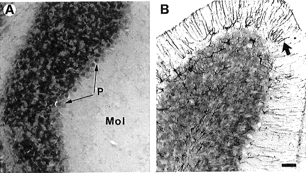
Fig. 3.
Astrocytes in the NPC mouse cerebellum as revealed by immunostaining for GFAP. Tissues were immunostanined for GFAP (black) and counterstained with creayl violet. A: In the NPC−/− mouse, there is no GFAP immunostaining. B: In the NPC−/− mouse, GFAP immunostaining is intense within the astrocytic fibers radiating from the Purkinjo cell layer up to the surface of the molecular layer (arrow points to one of many such glial fibers). Note the few astrocyte somata in the Purkinje cell layer. Abbreviations: Mol, molecular layer; P, Purkinje cell layer. Scale bar = 30 µm.

