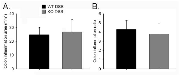
Figure 5. Colon Inflammation.
Inflammatory lesions such as ulcers and regions with thickened mucosa were measured in resected colons. Shown are plots for A. total inflamed colon area and B. colon inflammation ratio (inflamed area/total colon area). No significant differences were found between WT DSS (n=14) and KO DSS groups (n=11).

