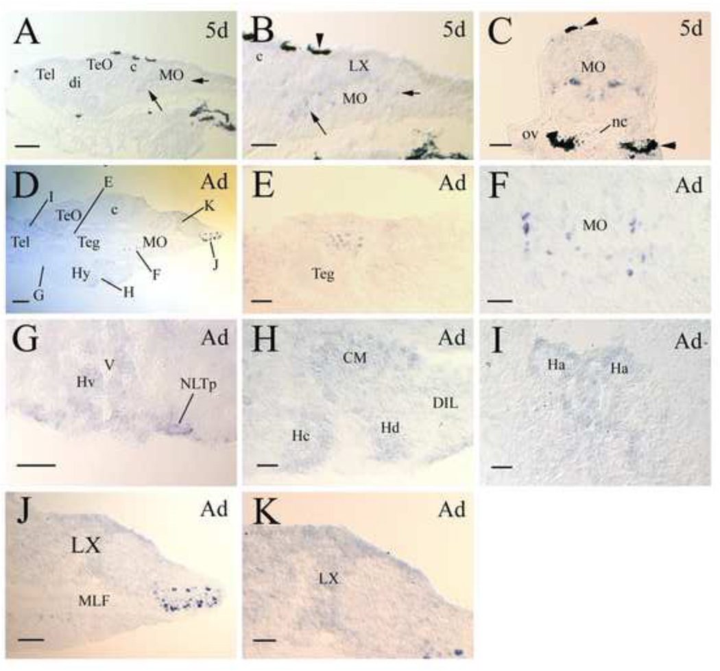
Figure 4.
lepr expression in developing and adult zebrafish brains. All panels show tissue sections, with anterior to the left and dorsal up for all sagittal sections, and dorsal up for all cross sections. Panel A shows a sagittal brain section from a 5-day old larva. Panel B is a higher magnification of the medulla oblongata (MO) shown in panel A. The short and long arrows in these two panels indicate the same respective regions with lepr expressing cells. The arrowhead in panel B points to a piece of pigmented epithelium. Panel C is a cross section of the MO at the otic vesicle (ov) level. Arrowheads indicate pigmented tissues. Panel D is a sagittal section showing the posterior telencephalon (Tel) and the rest of the brain. Letters in panel D refer to the area of this section shown in higher magnification in subsequent panels (E-I). Panels E, F, H, J and K are from sagittal sections, while panels G and I are from cross sections. Other abbreviations: c, cerebellum; CM, corpus mamillare; di, diencephalon; DIL, diffuse nucleus of the inferior lobe; Ha, habenula; Hc, caudal zone of periventricular hypothalamus; Hd, dorsal zone of periventricular hypothalamus; Hv, ventral zone of periventricular hypothalamus; Hy, hypothalamus; LX, vagal lobe; MLF, medial longitudinal fascicle; nc, notochord; NLTp, posterior part of the lateral tuberal nucleus; Teg, tegmentum; TeO, tectum opticum; V, ventricle. Scale bar = 100 µm for panels A and J, 200 µm for panel D, and 50 µm for the remaining panels.

