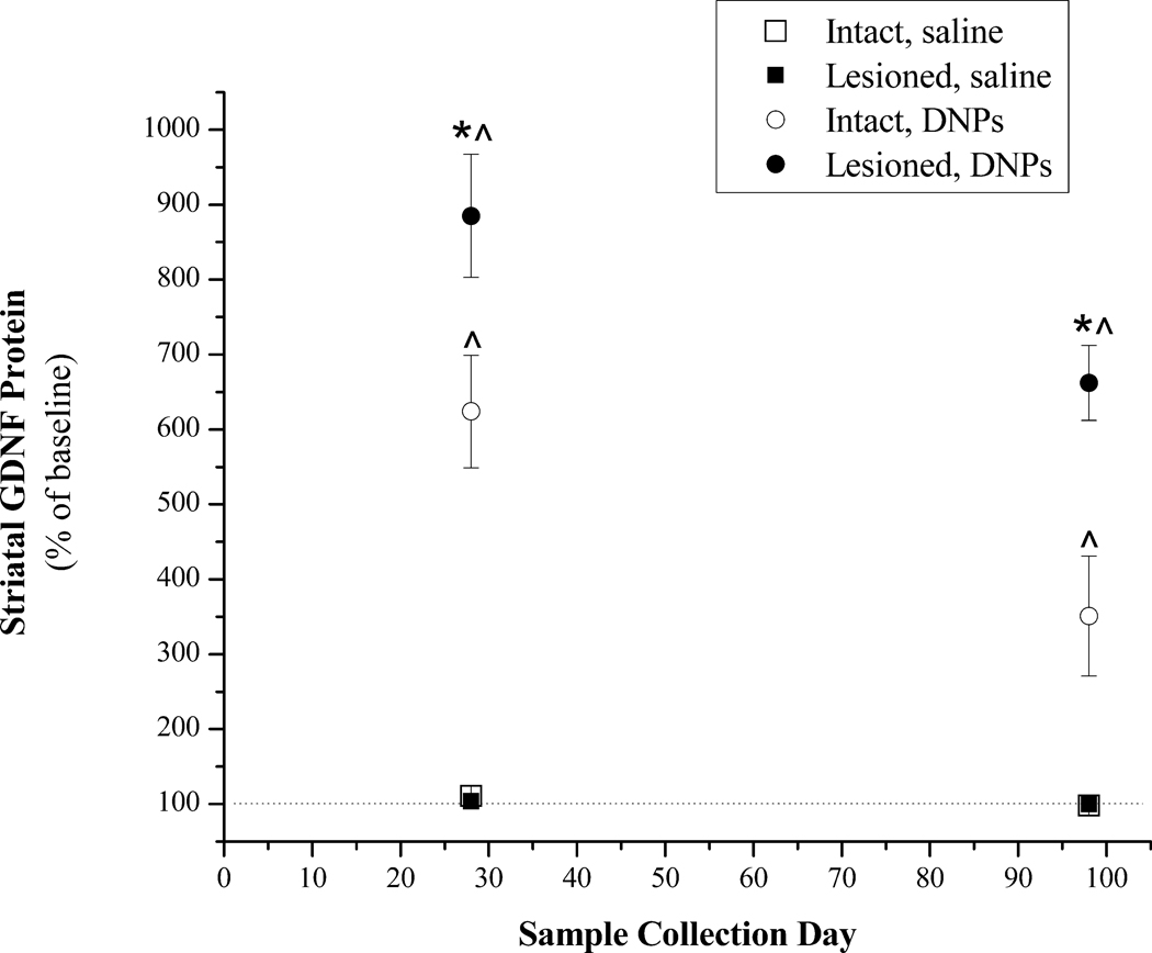
Figure 5.
Striatal GDNF protein levels at 28 or 98 days following a single injection of pGDNF_1b DNPs or saline into the intact or lesioned striatum. This study used naïve rats or rats with unilateral 6-OHDA lesions. Saline (8.0 µl) was injected into the left striatum of naïve rats (Intact, saline; n=5) or into the denervated striatum 4 weeks after 6-OHDA treatment (Lesioned, saline; n=5). pGDNF_1b DNPs (16.0 µg) was injected into the left striatum of naïve rats (Intact, DNPs; n=5) or into the denervated striatum 4 weeks after 6-OHDA treatment (Lesioned, DNPs; n=5). Animals were euthanized either 28 or 98 days following the saline or DNP injection and the injected and non-injected striatum were dissected from the brain. In each dissected sample GDNF protein content was determined by ELISA and data are graphed as a percent of baseline; that is, GDNF protein values for the injected side were divided by the values for the non-injected side (baseline). * p<0.05 Lesioned, DNPs vs. Intact, DNPs at 28 and 98 days; ^p<0.05 Lesioned, DN Ps or Intact, DNPs vs. Intact, saline or Lesioned, saline at 28 or 98 days.

