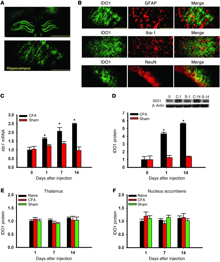
Figure 2. IDO1 expression in the hippocampus.
(A) IDO1 immunoreactivity was detected in the hippocampus. Scale bars: 1.0 mm (top), 50 μm (bottom). (B) Photomicrographs of colocalization between IDO1 and GFAP, Iba-1, or NeuN in the hippocampus. Scale bars: 50 μm. (C and D) Ido1 mRNA (C) and protein (D) expression was increased in the contralateral hippocampus of Wistar rats injected with CFA as detected by real-time PCR (C) and Western blot analysis (D). Day 0, baseline (naive rats); C-1 and C-14, samples taken on day 1 and day 14 from rats with CFA-induced arthritis; S-1 and S-14, samples taken on day 1 and day 14 from sham control rats. β-Actin was used as loading control. y axis shows fold change in IDO1 mRNA and protein expression. Mean ± SEM, n = 6–10, *P < 0.05 compared with sham control. (E and F) IDO1 expression (Western blot) was not upregulated in the thalamus (E) or nucleus accumbens (F) in Wistar rats with CFA-induced arthritis; n = 6, P > 0.05.

