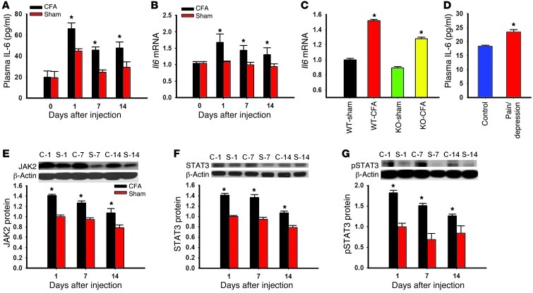
Figure 7. Role of IL-6 in the hippocampal JAK/STAT pathway.
(A and B) The plasma IL-6 concentration was increased (A, ELISA), as was Il6 mRNA expression in the hippocampus (B, real-time PCR), in rats with CFA-induced arthritis. (C) Il6 mRNA expression was also increased in the hippocampus of both IDO-knockout and wild-type mice with CFA-induced arthritis. Mean ± SEM, n = 6, *P < 0.05 compared with sham control. (D) The plasma IL-6 level was elevated in patients with both chronic back pain and depression (ELISA). Mean ± SEM, n = 13–20, *P < 0.05 compared with healthy control. (E–G) The expression of JAK2 (E), STAT3 (F), and p-STAT3 (G) in the hippocampus was increased in rats with CFA-induced arthritis (Western blot analysis). C-1, C-7, and C-14, samples taken on days 1, 7, and 14 from rats with CFA-induced arthritis; S-1, S-7, and S-14, samples taken on days 1, 7, and 14 from sham control rats. β-Actin was used as loading control. Mean ± SEM, n = 4–5, *P < 0.05 compared with sham control.

