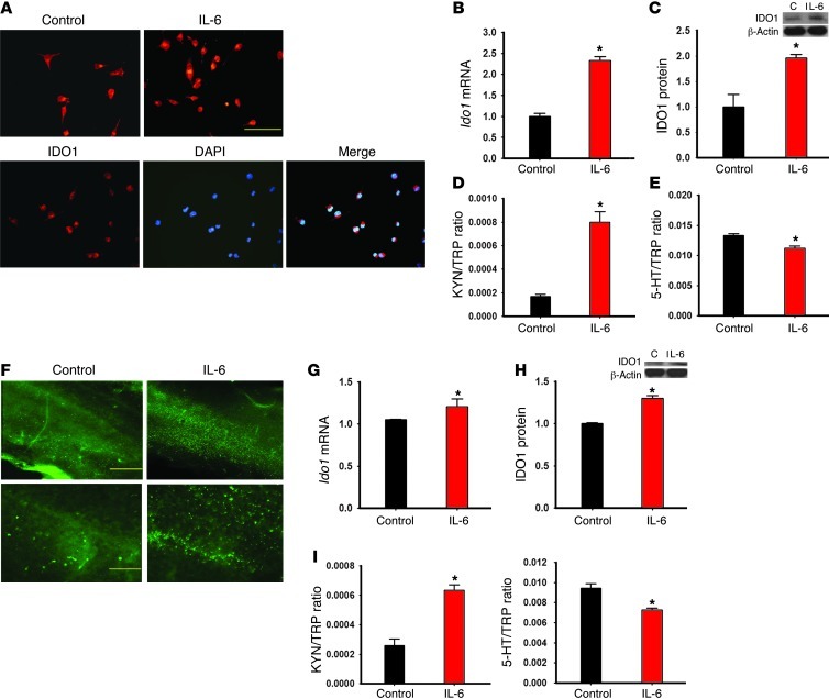
Figure 8. Effects of IL-6 on IDO1 expression in Neuro2a cells and organotypic hippocampal culture.
(A) IDO1 immunoreactivity was increased in IL-6–treated Neuro2a cells, as expressed in perinuclear cytoplasm when samples were costained with DAPI. Scale bar: 50 μm. (B–E) IDO1 mRNA (B) and protein (C) expression, as well as IDO activity (KYN/TRP ratio [D] and 5-HT/TRP ratio [E]), was increased in cultured Neuro2a cells after addition of IL-6 (0.5 ng/ml) for 24 hours. (F–H) Exposure of exogenous IL-6 (100 ng/ml) for 24 hours increased the expression of IDO1 (F: immunoreactivity; G: mRNA; H: Western blot) in hippocampal organotypic slice culture. Scale bars: 500 μm (top row) and 50 μm (bottom row). C, control. In addition, adding IL-6 for 24 hours also increased the KYN/TRP ratio and decreased the 5-HT/TRP ratio (HPLC) in the culture medium (I). *P < 0.05 compared with vehicle control.

