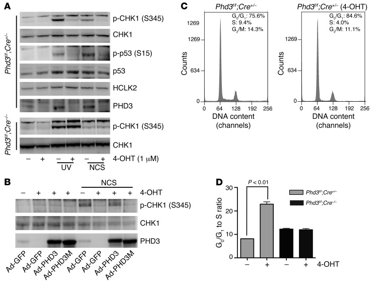
Figure 6. PHD3 plays a crucial role in the activation of the ATR/CHK1/p53 pathway following DNA damage and blocks G0/G1 to S phase transition in primary MEFs.
(A) After 3 days of treatment with 4-OHT, Phd3Β/Β;Cre+/– or Phd3Β/Β;Cre–/– MEFs were exposed to UV light (250 j/m2) or NCS (200 ng/ml) as indicated. Two hours later, cells were harvested, and Western blots were performed with the indicated antibodies. Representative blots from 3 similar experiments are shown. (B) Phd3Β/Β;Cre+/– MEFs were treated with 4-OHT for 3 days. On the second day, cells were infected with Ad-PHD3 or Ad-PHD3M as indicated. Cells were then treated with NCS (200 ng/ml) for 2 hours, and cell lysates were analyzed by Western blot with the indicated antibodies. Representative experiments from 2 experiments are shown. (C and D) Phd3Β/Β;Cre+/– or Phd3Β/Β;Cre–/– MEFs were treated with 4-OHT (1 μM) or solvent only for 3 days. Cells were then stained with propidium iodide (PI) and analyzed with FACS. The representative histograms of Phd3Β/Β;Cre+/– MEFs treated with or without 4-OHT are shown in C. (D) The relative sizes of G0/G1 to S phase ratio of the indicated MEFs (mean ± SEM). n = 3; *P < 0.01.

