
Figure 1.
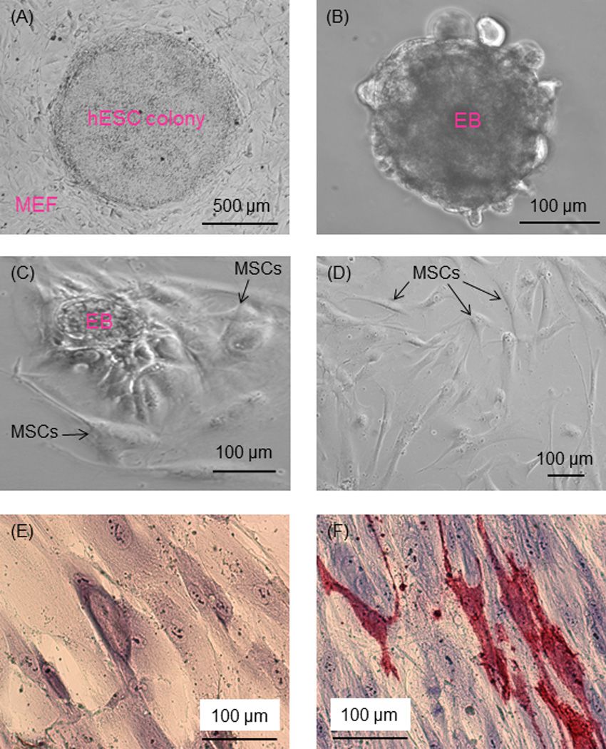
Phase-contrast images of hESC culture. (A) hESC colonies were cultured on MEF feeder layer. (B) hESC colonies were dissociated into clumps, which formed EBs in suspension culture. (C) In further culture of EBs, cells migrated out of the EBs. (D) The outgrowth of cells had a morphology similar to MSCs (passage 3 shown in the example in D). (E, F) Passage 4 cells were cultured in MSC growth medium or osteogenic medium for 21 d. Cells in growth medium had no ALP staining (E). Cells in osteogenic medium had ALP staining (F), where red color represents ALP-positive cells.