
Figure 2.
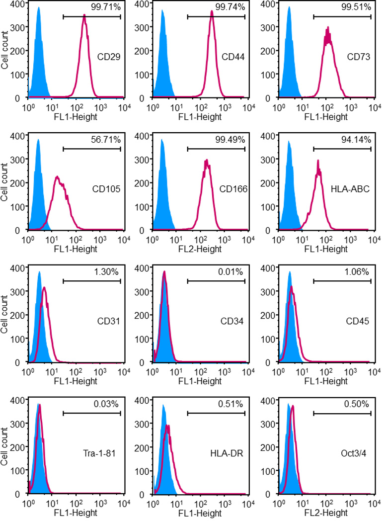
Immunophenotyping of the hESCd-MSCs. Flow cytometry showed that hESCd-MSCs expressed a number of cell surface markers characteristic of MSCs, and were negative for typical hematopoietic and endothelial cell markers. For example, MSC surface markers CD29, CD44, CD73, and CD166 were expressed to levels greater than 99.4%, while expressions of hematopoietic markers (CD31, CD34, CD45) were less than 1.5%.