
Figure 6.
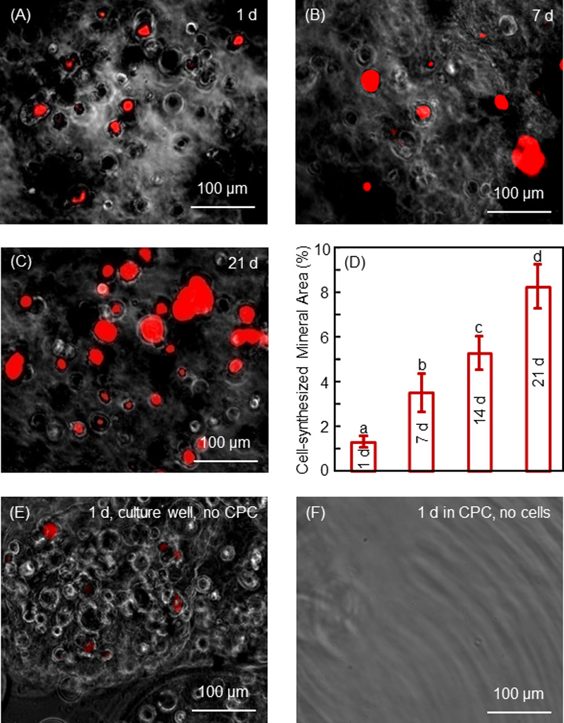
Mineral synthesis by hESC-MSCs in hydrogel microbeads in CPC. Xylenol stained minerals into a red color for (A–C) 1 d, 7 d and 21 d, respectively. (D) The percentage of mineralization area stained in red. Each value is mean ± sd (n = 6). Values with dissimilar letters are significantly different from each other (p < 0.05). Mineral synthesis by the encapsulated hESCd-MSCs was increased by 7-fold in 21 d. (E) Microbeads with cell encapsulation were cultured for 1 d in cell culture well without CPC. (F) Microbeads without cells were placed in CPC for 1 d. The microbeads were then collected for xylenol staining. These results confirmed that the red staining in A–C was from the cells and not from CPC.