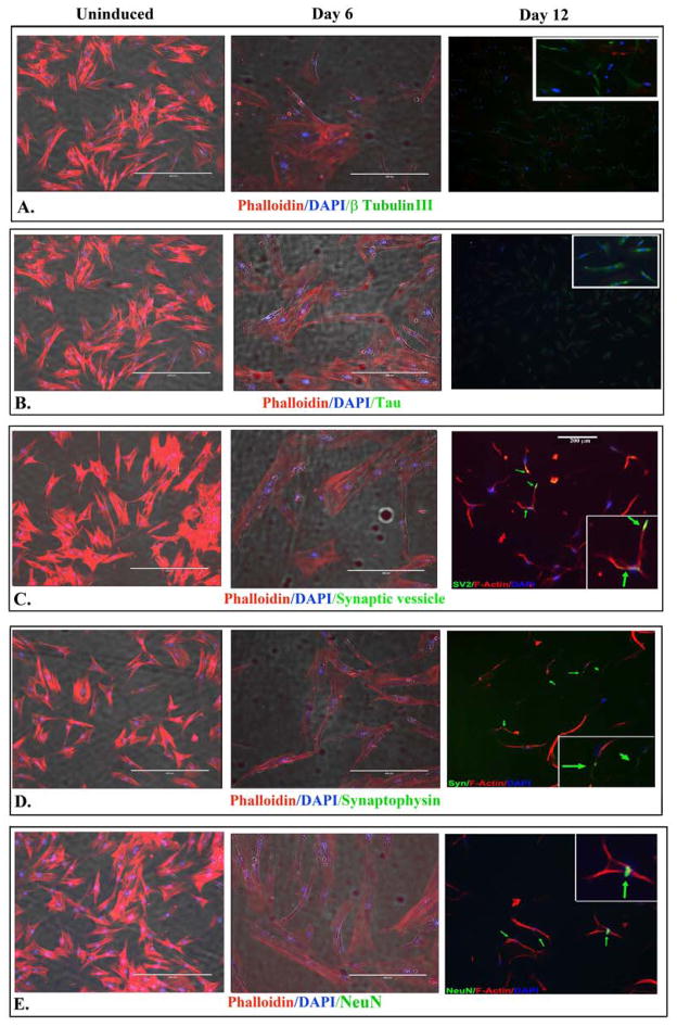
Fig 4. Immunofluorescence labeling for neuronal markers.
Shown are representative images (100x) of three different experiments for the following: β-tubulin III (A); Tau (B); Synaptic vesicle (C); Synaptophysin (D) and NeuN (E). The labelings were performed for uninduced MSCs and cells induced for 6 and 12 days. Co-labelings were performed with DAPI (blue) and phalloidin (red).

