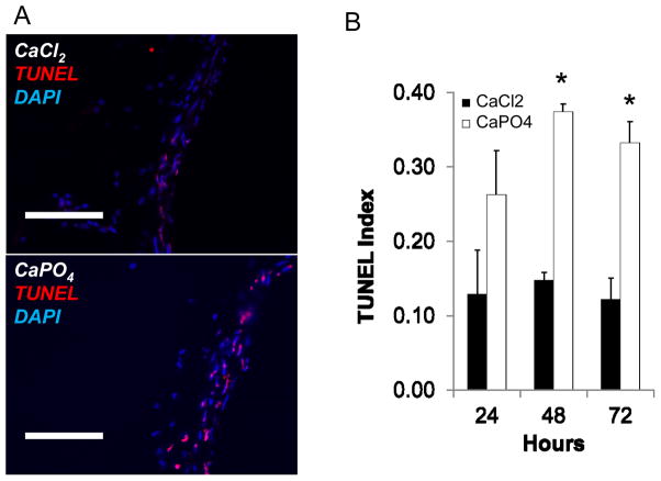
Figure 3. CaPO4-induced aneurysm displays apoptosis.
Mice were subjected to AAA induction with CaCl2 or CaPO4 and were sacrificed at indicated time points as described in Materials and Methods. A) Representative images of immunohistochemistry for apoptosis, as measured by TUNEL (red), nuclei shown by DAPI stain (blue) in treated arteries 3 days after injury. Scale bar = 100 μm. B) TUNEL index as determined by TUNEL positive cells/nuclei. Measurements taken from CaCl2- (black bar, ■CaCl2) and CaPO4 –treated (white bars, □CaPO4) arteries harvested 24, 48 or 72 hours after surgery. *p < 0.05, n = 3.

