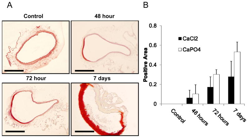
Figure 5. CaPO4-induced aneurysm samples contain medial calcification.
A) Arterial sections stained with Alizarin Red for calcium deposit detection, calcium appearing red on a pink and yellow background. ‘Control’ sections harvested from animals treated with NaCl only; ‘48 hour’,’ 72 hour’, and ‘7 day’ sections harvested at respective times after treatment by CaPO4 surgery. Scale bar = 200 μm for all images. B) Quantification of calcium content in arterial sections harvested from arteries with the conventional CaCl2 model (CaCl2) or the CaPO4 model (CaPO4). Data was expressed as a ratio of the total calcified media divided by the total medial area of each arterial section. n=4.

