
Figure 2. MCT1 required for neuronal survival in vitro.
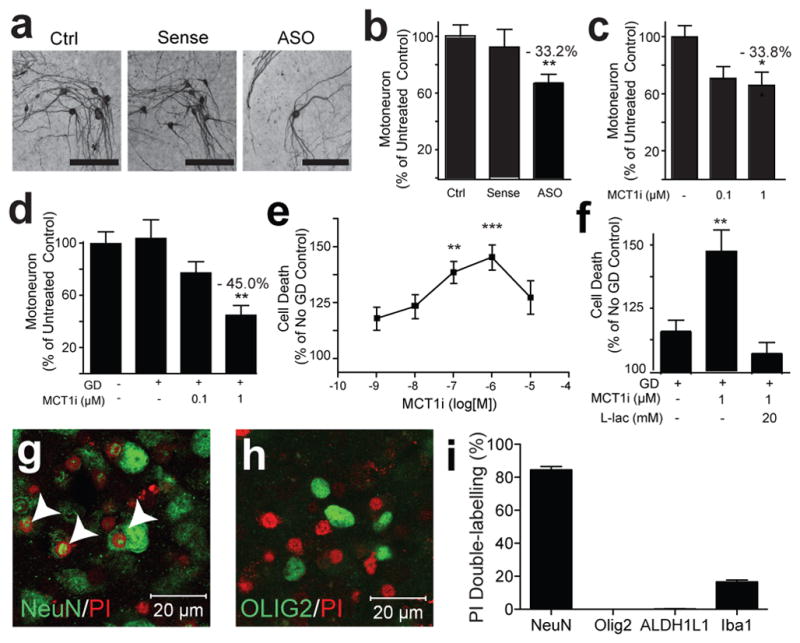
(a–d) Photomicrograph (a) and quantification of motoneurons in spinal cord slice cultures treated with media only (Ctrl), MCT1 sense oligonucleotides (Sense), or MCT1 antisense oligonucleotides (ASO) for 3 weeks (b; n ≥ 55 sections per group), following 3 weeks treatment with MCT1i (c; n ≥ 27 sections per group), or 2 hrs of glucose deprivation (GD) ± MCT1i (d; n ≥ 30 sections per group). (e,f) Propidium iodide (PI) uptake in slice cultures treated with 2 hrs GD + MCT1i (e; n ≥ 10 sections per group) or 2 hrs GD, ± MCT1i, ± 20 mM lactate (f; n = 15 for all groups). (g,h) PI uptake in slices treated with GD + MCT1i labelled with neuronal (g; co-localized cells marked with arrowheads) or oligodendroglia (h) marker. (i) Percentage of PI-labelled cells co-localizing cell-specific markers (n 10 sections per group, Error bars = ± S.E.M. *p < 0.05, **p < 0.01, ***p < 0.001).