
Figure 4. MCT1 heterozygote null mice develop widespread axonopathy.
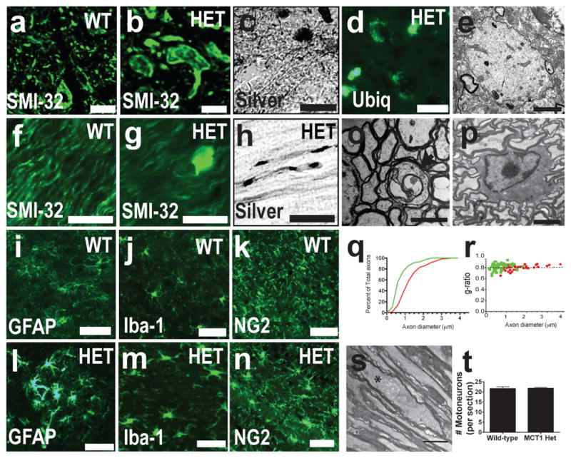
(a–d) Spinal cords of wild-type (WT) and MCT1+/− mice (HET) immunostained for non-phosporylated neurofilament (SMI-32) (a,b), silver stained (c), or immunostained for ubiquitin (d; Scale bars = 20 um). (e) Electron microscopy of axonal spheroid in spinal cord (scale bar = 2 μm). (f–h) Optic nerves of WT and HET mice immunostained for SMI-32 (f,g) or silver stained (h; scale bars = 20 um). (i–n) GFAP (i.l), Iba-1 (j,m), and NG2 (k,n) in WT (i–k) and HET mice (l–n ; Scale bars = 50 um). (o,p) Degenerating axon (o; indicated by arrow, scale bar = 2 μm) and intact oligodendrocyte (p; scale bar = 6 μm) in optic nerve. (q,r) Axon diameter (q) and g ratio (r) in HET (green line/dots ; n=158) and WT (red line/dots ; n=78) mice. (s) Distended mitochondrion (astrerisk) in HET optic nerve. (t) Number of spinal motorneurons per section from WT and HET mice.