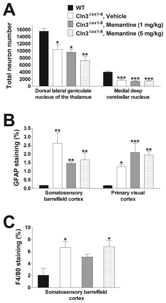
Fig. 3. Acute treatment with memantine has no effect on the neuropathological changes in 6-month-old Cln3Δex1-6 mice.
Six-month-old Cln3Δex1-6 mice acutely treated with memantine (1 or 5 mg/kg) or the vehicle of memantine (sterile 0.9% NaCl) were perfusion-fixed 8 days after the treatment (4 mice from each treatment group), and their brains were histologically analyzed. Four 6-month-old, untreated WT mice were also perfusion-fixed, and their brains were histologically analyzed, as well. (A) Localized neuronal loss in the thalamus and cerebellum. To survey the survival of neuron populations that are vulnerable in Cln3Δex1-6 mice, counts of the large projection neurons in the thalamus (dorsal lateral geniculate nucleus) and in the medial deep cerebellar nucleus were made. These counts were obtained using the StereoInvestigator software. (B) Astrocytosis in the cortex. Quantitative determination of astrocytosis in the somatosensory barrelfield cortex and the primary visual cortex was performed by thresholding image analysis following immunohistochemical staining for the astrocytic marker, GFAP. (C) Microgliosis in the cerebellum. Quantitative determination of microgliosis in the somatosensory barrelfield cortex and the primary visual cortex was performed by thresholding image analysis following immunohistochemical staining for the microglial marker F4/80.
Statistical significance was calculated by one-way ANOVA with Bonferroni’s post-test: *p<0.05, **p<0.01 and ***p<0.001 as compared to WT.

