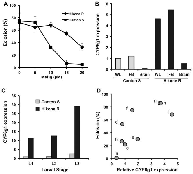
Fig. 1.
MeHg tolerance and CYP6g1 expression in wild strains of flies. (A) Tolerance to MeHg during development in the Canton S and Hikone R wild strains is determined by eclosion assays. Greater rates of eclosion are seen for Hikone R strain on 10–20 μM MeHg food than for Canton S. (B) Expression levels of CYP6g1 transcripts are determined using total RNA from extracts of whole larvae (WL), fat body (FB) or central nervous system (Brain) of 3rd instar larvae of the Canton S and Hikone R strains. Determinations are done by qRT-PCR and expression levels normalized to Canton S strain WL. (C) Relative expression level of CYP6g1 transcripts across larval developmental stages are determined by qRT-PCR using total RNA from extracts of whole larvae of Canton S and Hikone R strains. Expression levels are normalized to Cantons 1st instar larvae (L1, L2, L3 = 1st, 2nd, 3rd instar larvae, respectively). (D) A comparison of tolerance to MeHg and CYP6g1 expression level is determined in nine wild strains of flies previously reported to have various levels of CYP6g1 (Daborn et al., 2002). Wild strains are analyzed by comparisons of CYP6g1expression (qRT-PCR, normalized to Canton S) and MeHg tolerance determined by eclosion rates on 10 μM MeHg food (Wild strains are: (a) VAG1; (b) Swedish-C; (c) Canton S; (d) PVM; (e) Wild 5A; (f) Reids 3; (g) Hikone R; (h) Wild 1B; (i) Wild 5C).

