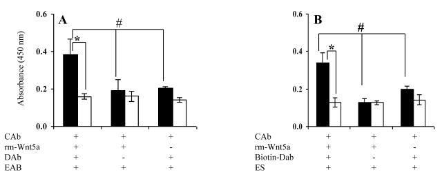
Figure 6.
PEG affords detection of rm-Wnt5a in the sandwich ELISA. Rabbit anti-human Wnt5a served as the capture antibody (CAb) in both (A) and (B). Goat anti-mouse Wnt5a (DAb) and HRP conjugated F(ab’)2 donkey anti-goat IgG (EAb) detection system was used in (A), and biotinylated goat anti-mouse Wnt5a and HRP-streptavidin detection system was used in (B). Black bar indicates the presence of PEG during the rm-Wnt5a and detection antibody binding stages of the ELISA, white bar indicates no PEG. Concentration of rm-Wnt5a was 1 μg/ml. OD was measured after 2 hrs of enzyme-substrate reaction. * p≤0.05 via Student’s t-test # p≤0.05 via Tukey-Kramer. All values are mean ± SD of triplicates.

