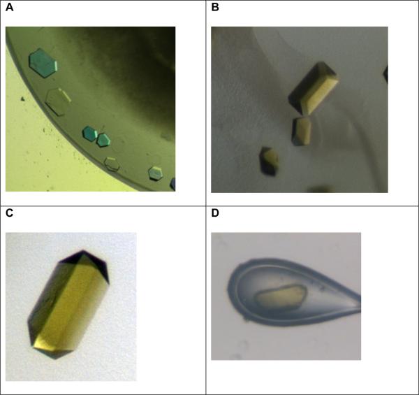
Figure 3. Gallery of crystals of the c-ring of the ATP synthase.
A: Initial small crystals of the c14-ring. The crystals are very thin plates (diameter 50 μM, height less than 10 μM). They were grown from a starting solution that contained the intact ATP synthase. Please note that the picture is taken under polarized light and therefore appear with false colors.
B, C and D: These pictures were taken under white light without polarization filter to show the true colors of the crystals.
B: Improved crystals of the c-ring (diameter 100 μM, height 50 μM), showing a strong yellow color. The crystals were grown from a starting solution that contains the intact ATP synthase.
C and D: Crystals of the isolated c-ring of the ATP synthase. Please note that the crystals show the same strong yellow color as the crystals grown from the intact enzyme.

