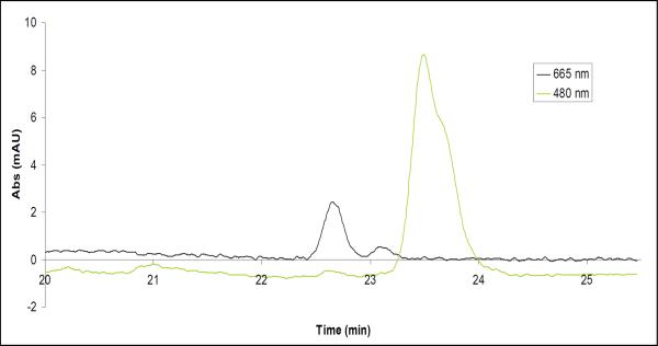
Figure 6. Reverse-phase HPLC analysis of the pigment content of the c-ring crystals.
Protein crystals were briefly subjected to a solution of 80% acetone/20% water to denature the protein and extract the pigments. The solution was then separated using a reverse-phase HPLC (as described in Methods) and two major peaks were observed. The peak observed by the 665 nm detector was identified as chlorophyll and pheophytin by spectral analysis. The peak observed by the 480 nm detector was identified as beta-carotene by spectral analysis.

