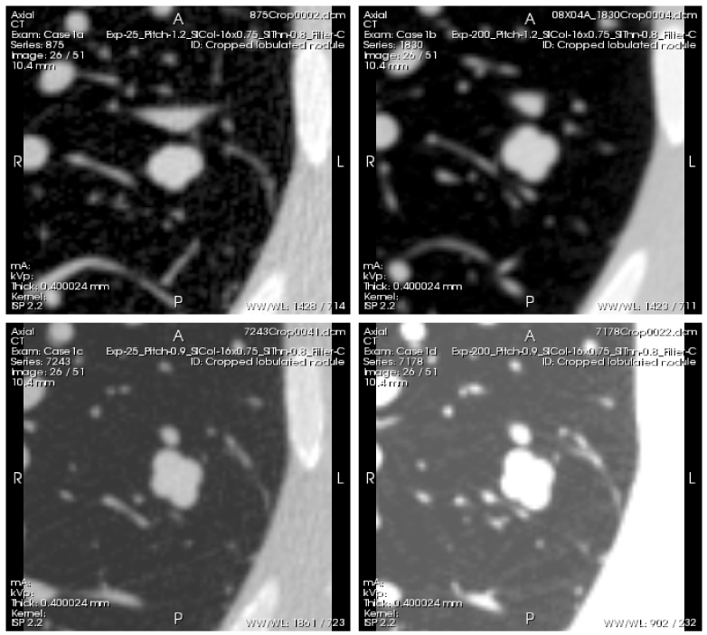
Fig. 4.

Example scans of a lobulated nodule with −10HU density and 10mm diameter (nominal volume equal to that of a 10 mm diameter sphere), acquired with the following 4 different protocols: Top left (Case 1a)- low exposure (25 mAs), 1.2 pitch. Top right (Case 1b)- high exposure (200 mAs), 1.2 pitch. Bottom left (Case 1c)- low exposure (25m As), 0.9 pitch. Bottom right (Case 1d)- high exposure (200m As), 0.9 pitch. Reconstructed slice thickness was 0.8 mm for all scans. The series of scans can be viewed by clicking on the Interactive Science Publishing (ISP) hyperlink ( View 1).
