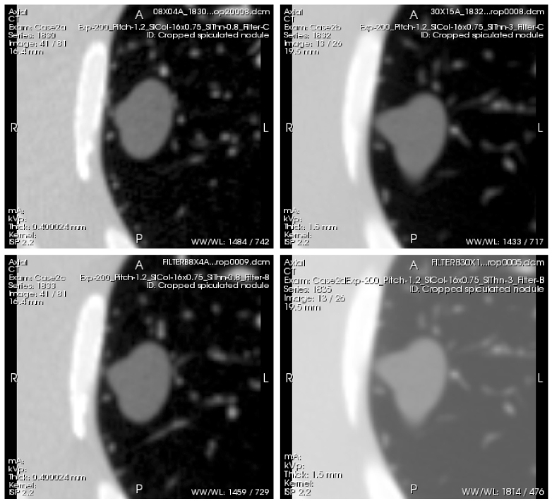
Fig. 5.

Example scans of a spiculated nodule of −630HU density and 20 mm diameter (nominal volume equal to that of a 20 mm diameter sphere), acquired with the following 4 protocols: Top left (Case 2a)- thin slice thickness (0.8 mm), detail reconstruction kernel (BF60). Top right (Case 2b)- thick slice thickness (3.0mm), detail reconstruction kernel (BF60). Bottom left (Case 2c)- thin slice thickness (0.8 mm), medium reconstruction kernel (BF40). Bottom right (Case 2d)- thick slice thickness (3.0 mm), medium reconstruction kernel (BF40). All scans were acquired with a high exposure (200 mAs) and 1.2 pitch. The whole series of each scan can be viewed by clicking on the Interactive Science Publishing (ISP) hyperlink ( View 2).
