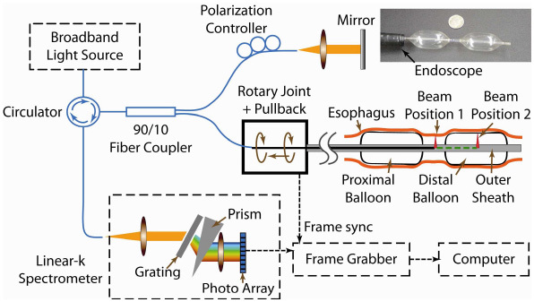
Fig. 1.

System schematic of the double-balloon-based SD-EOCT system. Within the catheter, the beam can be located between two balloons for imaging without balloon-tissue contact (labeled beam position 1) or in a balloon for imaging with contact (labeled beam position 2). The inset photo (top right) shows the double-balloon catheter (compared with a dime) inserted through the GI endoscope and inflated.
