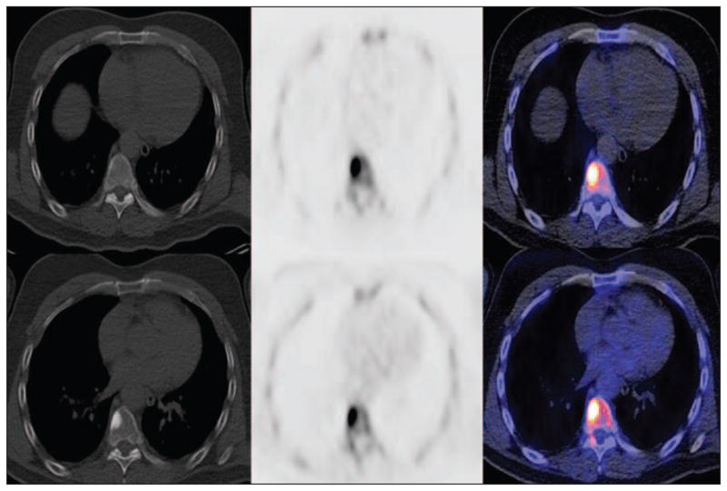
Fig. 5.

54-year-old man with biochemical recurrence of prostate cancer and negative conventional bone scintigraphy. CT (left), 18F-NaF PET (middle), and fused 18F-NaF PET/CT (right) transaxial images show subtle sclerosis at right anterolateral aspect of thoracic vertebral body, which is clearly active on PET before androgen deprivation therapy (top) and remains active with increasing sclerosis after 4 months of treatment (bottom).
