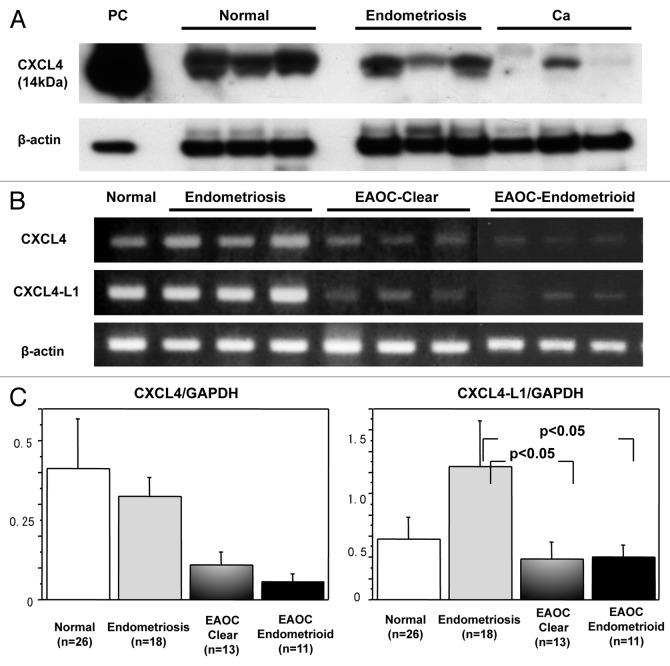
Figure 1. Expression of CXCL4 in normal, endometriosis and cancer tissues. (A) western blot analysis is performed using anti-CXCL4 antibody that recognizes both CXCL4 and CXCL4-L1. Representative cases of normal, endometriosis and cancer tissues are shown. β-actin is used as a control. Platelet-derived protein is used as positive control. (B) Results of RT-PCR analysis are shown. CXCL4-specific and CXCL4-L1-specific primers are used, respectively. The PCR product samples were subjected to 2% agarose gel electrophoresis and visualized by staining with ethidium bromide. (C) Real-time RT-PCR was performed and the expression levels of CXCL4 (left graph) and CXCL4-L1 (right graph) normalized by GAPDH were investigated among normal (n = 26), endometriosis (n = 18), clear cell cancer (n = 13) and endometrioid cancer (n = 11) tissues.

An official website of the United States government
Here's how you know
Official websites use .gov
A
.gov website belongs to an official
government organization in the United States.
Secure .gov websites use HTTPS
A lock (
) or https:// means you've safely
connected to the .gov website. Share sensitive
information only on official, secure websites.
