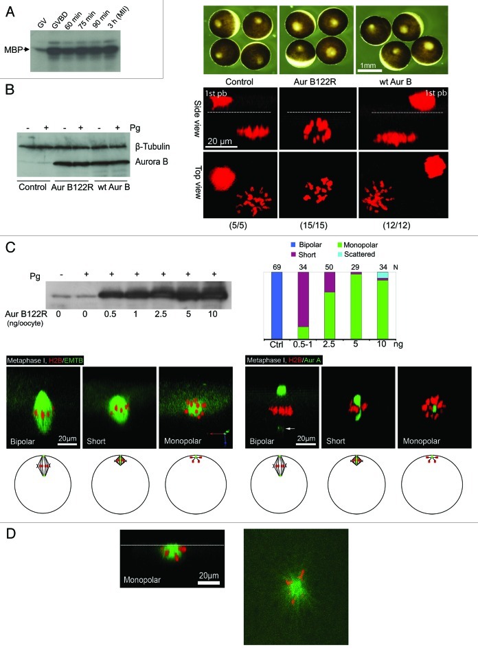
Figure 2. Aur-B122R interferes with spindle bipolarity in frog ocoytes. (A) Aur-B activation during first polar body emission: Oocytes were incubated with progesterone and monitored every 10 min for GVBD. GVBD oocytes were picked out and further incubated for the indicated period of time. Groups of 30 oocytes each were lysed and the extracts were subjected to immunoprecipitation with anti-Aur-B antibodies, followed by in vitro kinase assays using myelin basic protein (MBP) as substrate, as described in our previous publication.19 (B) Overexpression of Aur-B122R, but not WT Aur-B, inhibits polar body emission: Oocytes were injected with RFP-H2B mRNA (control), plus Aur-B122R mRNA (5 ng per oocyte) or WT Aur-B mRNA (5 ng per oocyte) and treated overnight with progesterone. Oocytes were imaged live and each of the representative oocytes is shown in both side view and top view (total numbers indicated). Dash lines denote plasma membrane. 1st pb = first polar body (trapped between the vitelline membrane and plasma membrane, See Fig. 1A). Also shown are bright field images of representative oocytes at time of confocal imaging, as well as a representative immunoblot depicting the levels of overexpression of WT Aur-B and Aur-B122R, compared with endogenous Aur-B (control). β-tubulin blotting served as internal control. (C) Phenotypes of Aur-B122R overexpression: Control oocytes and oocytes injected with the indicated amount of Aur-B122R mRNA, plus the indicated fluorescence probes. Individual oocytes were examined live between 90 and 110 min after GVBD and classified as bipolar (spindle), short (spindle), monopolar (spindle) and scattered (chromosomes). The graph summarizes 20 experiments, with typical images of the indicated phenotypes (“scattered” not shown). The left three images (H2B/EMTB) are 3-D views slightly tilted upward to show that all spindles (bipolar, short and monopolar) were attached to the cortex. Arrow points to Aur-A signal at the cytoplasmic pole. Also shown is a representative anti-Aur-B immunoblot of extracts of control oocytes or oocytes injected with the indicated amounts of Aur-B122R (+ or – progesterone stimulation). Five oocytes were lysed in each group but only 1/5 of the extracts (equivalent to one oocyte) were analyzed. The schematics depict the various phenotypes in intact oocytes (side view), with animal pole at top. Green: Aur-A (spindle pole); red: chromosomes; black lines: microtubules. (D) Surface view of a monopolar Aur-B122R oocyte: The left panel is a 3-D rendered image (side view) with a dash line depicting the oocyte surface. The right panel is a single plane confocal image of the oocyte surface, depicting chromosomes attaching to a central core of microtubules (presumably kinetochore microtubules) and radial polar microtubules growing beyond the chromosomes.

An official website of the United States government
Here's how you know
Official websites use .gov
A
.gov website belongs to an official
government organization in the United States.
Secure .gov websites use HTTPS
A lock (
) or https:// means you've safely
connected to the .gov website. Share sensitive
information only on official, secure websites.
