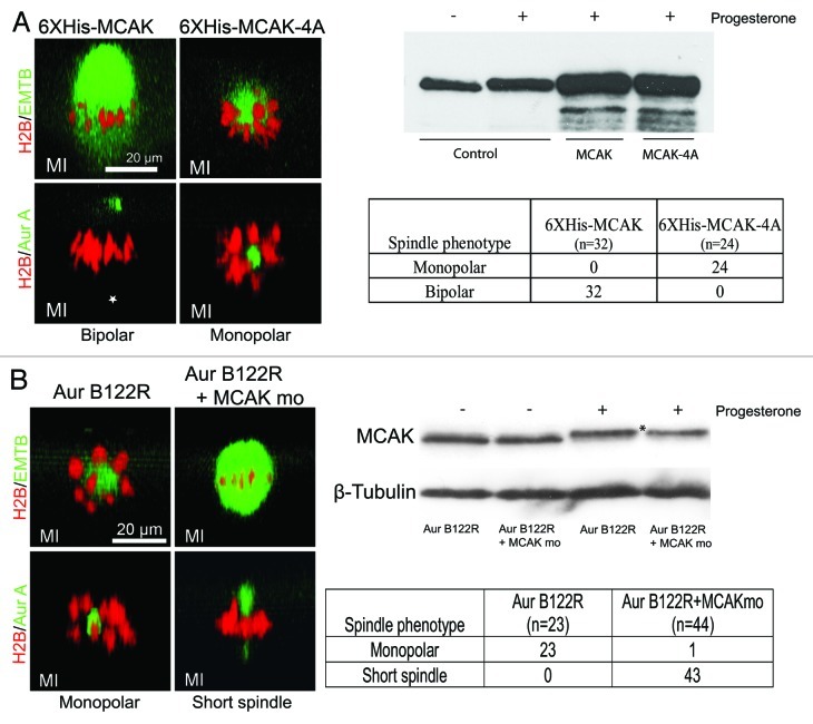
Figure 3. Aur-B regulates spindle bipolarity by suppressing MCAK. (A) MCAK-4A, but not WT MCAK, caused monopolar spindles in meiosis I: Oocytes were injected with RFP-H2B and eGFP-EMTB, or RFP-H2B and Alexa488 anti-Aur A, together with either 20 ng of 6XHis-MCAK or 20 ng of 6XHis-MCAK-4A. The oocytes were treated with progesterone and examined live between 90–110 min after GVBD (prometaphase to metaphase). Shown are typical confocal images (side view) of the bipolar spindles in MCAK oocytes and monopolar spindles in MCAK-4A oocytes, as summarized in the table. Note that the cytoplamic pole (*) of the control oocyte is not visible due to its depth and the relatively weak Aur-A signal. Also shown is a representative anti-MCAK immunoblot of extracts of oocytes injected with 20 ng of 6XHis-MCAK or 6XHis-MCAK-4A, compared with endogenous MCAK (control). Five oocytes were lysed in each group but only 1/5 of the extracts (equivalent to one oocyte) were analyzed. (B) MCAK mo rescued Aur-B122R monopolar phenotype: Oocytes were injected with Aur-B122R mRNA (10 ng per oocyte), together with the appropriate fluorescence probes (RFP-H2B, eGFP-EMTB or Alexa488 anti-Aur A). Half of the oocytes were further injected with MCAK mo (1 mM, 20 nL per oocyte). The oocytes were treated with progesterone and examined live between 90–110 min after GVBD (prometaphase to metaphase). Shown are typical confocal images (side view) of the monopolar spindles in Aur-B122R oocytes and short spindles in oocytes co-injected with Aur-B122R and MCAK mo, as summarized in the table. Also shown is a representative (out of a total of 5 independent experiments) anti-MCAK immunoblot of extracts of oocytes injected with Aur-B122R alone, or together with MCAK mo, following overnight incubation in the presence or absence of progesterone.

An official website of the United States government
Here's how you know
Official websites use .gov
A
.gov website belongs to an official
government organization in the United States.
Secure .gov websites use HTTPS
A lock (
) or https:// means you've safely
connected to the .gov website. Share sensitive
information only on official, secure websites.
