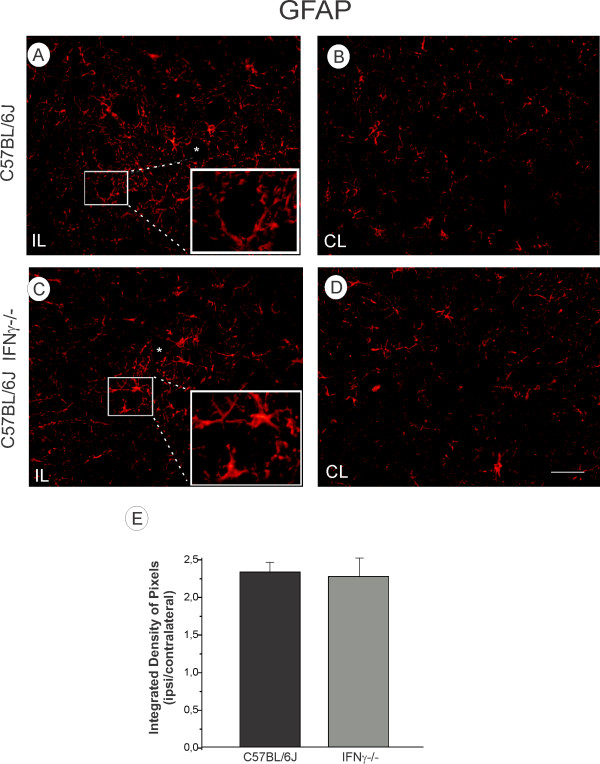
Figure 4.
(A, C) Ipsilateral side (IL, lesioned) of the C57BL/6J and IFNγ−/− mice, respectively. The lesion upregulated GFAP labelling in both strains. (B, D) Contralateral side (CL, unlesioned) of the lumbar spinal cord. Observe the presence of more hypertrophied astrocytes surrounding motoneurons in the IFNγ−/− mice (inset in A) as compared to C57BL/6J (inset in C). (F) Graph representing the ipsi/contralateral ratio (p > 0.05). Scale: 50 μm.

