Figure 1.
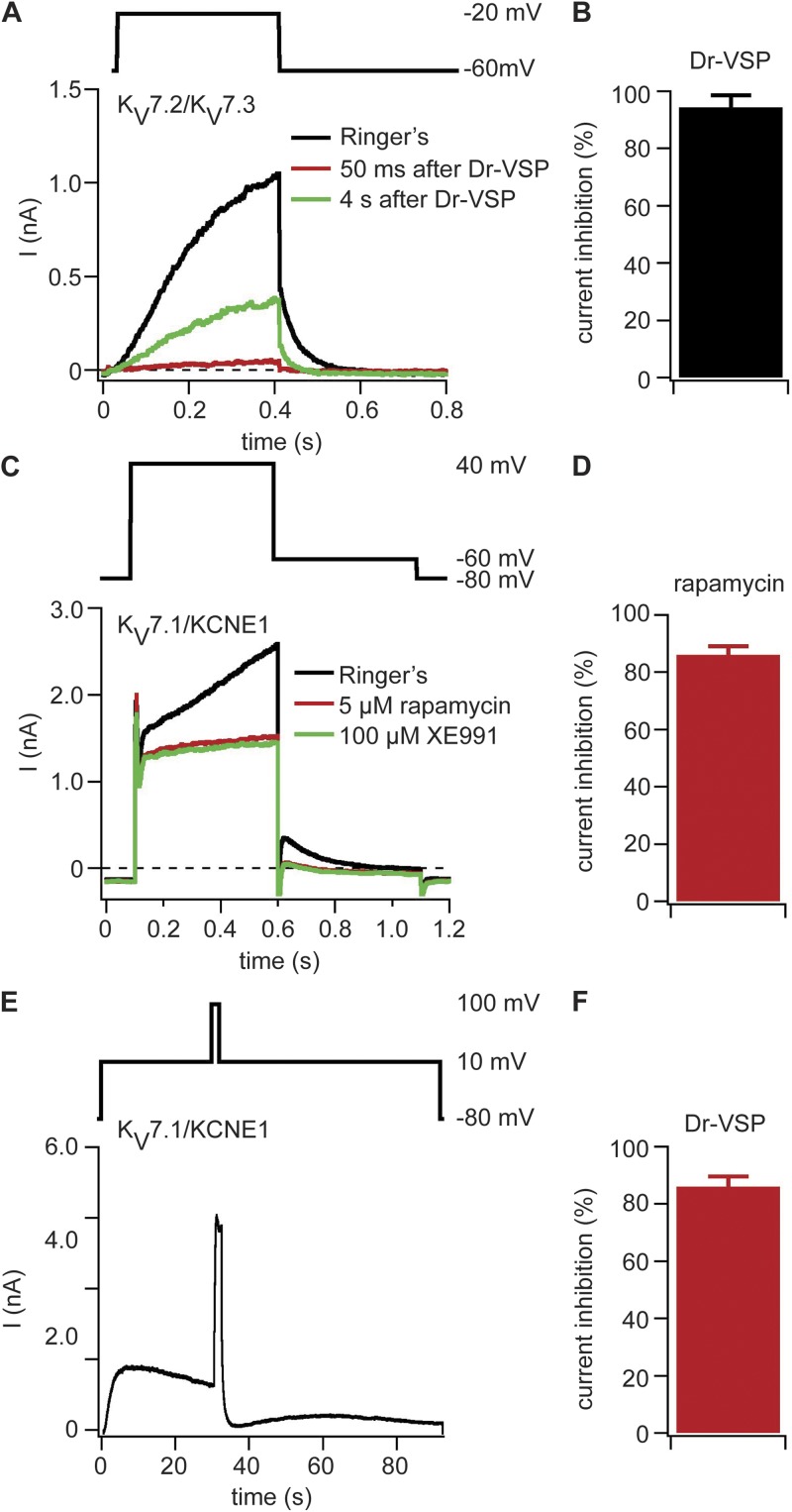
PI(4,5)P2 depletion at the plasma membrane inhibits KV7.x channels. (A) Currents with coexpression of KV7.2, KV7.3, and Dr-VSP in tsA-201 cells. The black current trace was recorded before Dr-VSP activation, the red trace was recorded 50 ms after Dr-VSP activation by a 2-s pulse to 100 mV, and the green trace was recorded 4 s after Dr-VSP activation. Test pulse protocol is shown above the traces. (B) Reduction of KV7.2/KV7.3-mediated tail currents by activation of Dr-VSP (n = 10). (C) Currents traces recorded from a cell expressing KV7.1, KCNE1, Ins-5-P-FKBP-CFP, and LDR-CFP. Traces are shown before application of 5 µM rapamycin (black), after 60 s of rapamycin application (red), and after application of 100 µM XE991 (green). (D) Rapamycin-induced inhibition of XE991-sensitive current (n = 5). (E) Current trace of a cell expressing KV7.1, KCNE1, and Dr-VSP. Currents were recorded at 10 mV, Dr-VSP was activated after 30 s with a 2-s long test pulse to 100 mV, and then the membrane potential was returned to 10 mV. At the end of the recording, XE991 was applied to determine the amount of KV7.1-mediated current (not depicted). (F) Inhibition of XE991-sensitive current by Dr-VSP activation (n = 6). (B, D, and F) Error bars represent ±SEM.

