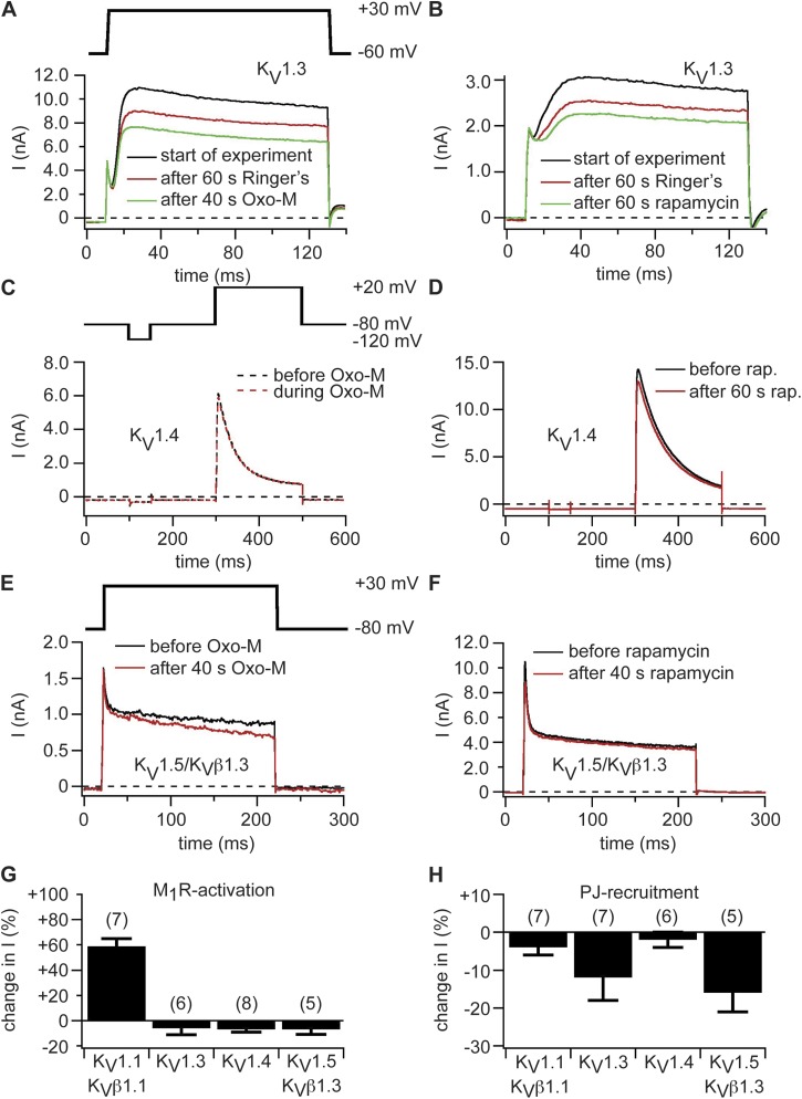
Figure 4.
KV1.3, KV1.4, and KV1.5 channels are insensitive to depletion of PI(4,5)P2 at the plasma membrane. (A) Current traces of KV1.3 channels coexpressed with M1R. Black indicates traces at start of experiment, red indicates after 60 s in Ringer’s, and green indicates traces after 40-s superfusion with 10 µM Oxo-M (100 s after start of experiment). (B) Currents in KV1.3 channels coexpressed with pseudojanin-YFP and LDR-CFP. Black indicates traces at start of experiment, red indicates after 60 s in Ringer’s, and green indicates traces after 60-s rapamycin application (120 s after start of experiment). Same pulse protocol as in A. (C) Currents in a cell expressing KV1.4 and M1R. Black indicates current trace in Ringer’s solution, and red indicates current trace during application of 10 µM Oxo-M. (D) KV1.4 channels coexpressed with pseudojanin-YFP and LDR-CFP. Black indicates current before application of rapamycin (rap.), and red indicates traces after 60 s of rapamycin application. (E and F) Currents in KV1.5 channels coexpressed with KVβ1.3 and M1R (E) or pseudojanin-YFP and LDR-CFP (F). Black indicates current before application of Oxo-M (E) or rapamycin (F), and red indicates current after 40-s application of Oxo-M (E) or rapamycin (F). (G and H) Percent changes in steady-state current amplitudes of KV1.x channels after activation of M1R (G) or recruitment of pseudojanin (PJ) to the plasma membrane (H). Numbers in parentheses indicate n numbers for individual experiments. Error bars represent ±SEM.

