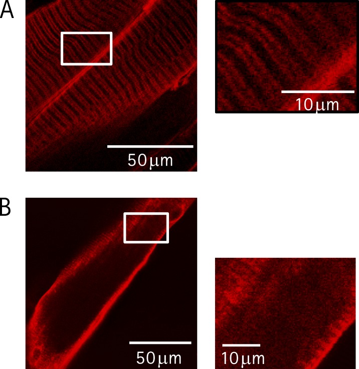
Figure 3.
TPLSM evaluation of TTS disruption by osmotic shock treatment. (A) TPLSM fluorescence image of an intact FDB muscle stained with di-8-ANEPPS. The inset is a threefold enlargement of the area defined by the white rectangle. (B) TPLSM fluorescence image of an isolated fiber stained with di-8-ANEPPS after being submitted to a formamide-based osmotic shock. In the area delimited by the rectangle, the plane of the TPLSM section was tangential to the fiber periphery. This area is enlarged threefold in the inset.

