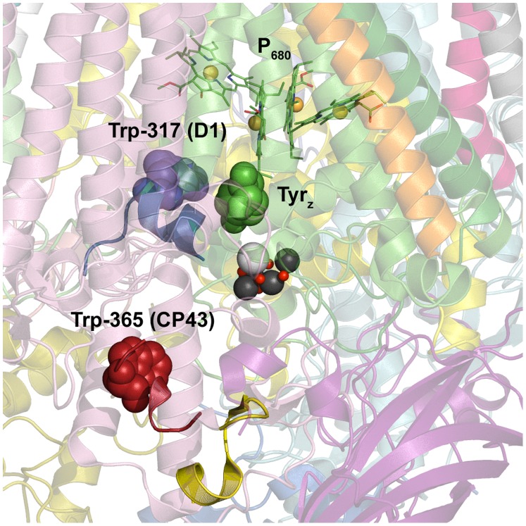
Figure 8. Predicted locations of NFK modifications, NFK365-CP43 and NFK317-D1, in the T. vulcanus PSII structure [2].
The OEC is shown in black, grey, and red. P680 and YZ (green spacefill) are shown above the OEC. The CP43 and D1 backbones are displayed in pink and green, respectively. The side chain of Trp-365 in CP43 is in red spacefill. The side chain of Trp-317 in D1 is in blue spacefill. MS/MS detected tryptic peptides corresponding to fraction A (red and yellow combined), B (blue), and C (red) are highlighted. The image was rendered with the Pymol Molecular Grapics System (www.pymol.org).

