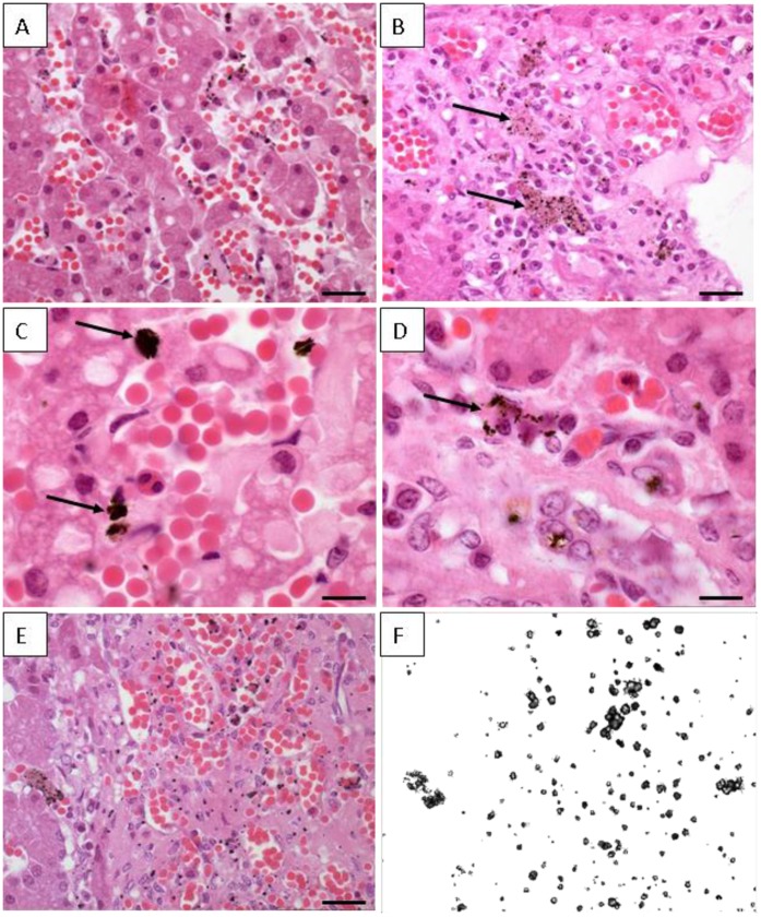
Figure 4. Histological sections from hepatic tissue of Guiana dolphin (Sotalia guianensis).
The sample is from a 9-year-old male dolphin from Guanabara Bay, Rio de Janeiro State. (A) Semi-thin section of the hepatic tissue showing the hepatocytes arranged in layers or plates; Longitudinal blood vessel filled with numerous red cells can be seen; Barr = 80 µm. (B) Different field of the organ showing the hepatic parenchyma with many blood vessel and some dark deposits within the Kupffer cells (arrows); Barr = 100 µm. (C) Agglomerated dark deposits found in Kupffer cells (arrows); Barr = 20 µm. (D) Deposits distributed in small dots (arrows); Barr = 20 µm. (E) Semi-thin section of dolphin liver showing the cells with dark inclusions; Barr = 100 µm. (F) - Same area depicted in figure E observed under polaryzed light; all the dark dots are birefringents materials and corresponds to deposits within the Kupffer cells.

