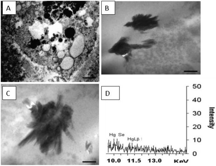
Figure 5. Ultrastructure of Guiana dolphin (Sotalia guianensis) liver.
The sample is from a 9-year-old male dolphin from Guanabara Bay, Rio de Janeiro State. (A) Electron microcopy image showing a Kupffer cell and its organelles. Numerous circular vesicles, some electron-denses, other electron-lucents; Barr = 5 µm. (B) Image of stick-like shaped electron-dense deposits found in Kupffer cells; Barr = 1 µm. (C) A detail of other electron-dense deposit; Barr = 500 nm. (D) X-ray microanalysis spectra showing the elements Hg and Se as components of those dark deposits found within Kupffer cells.

