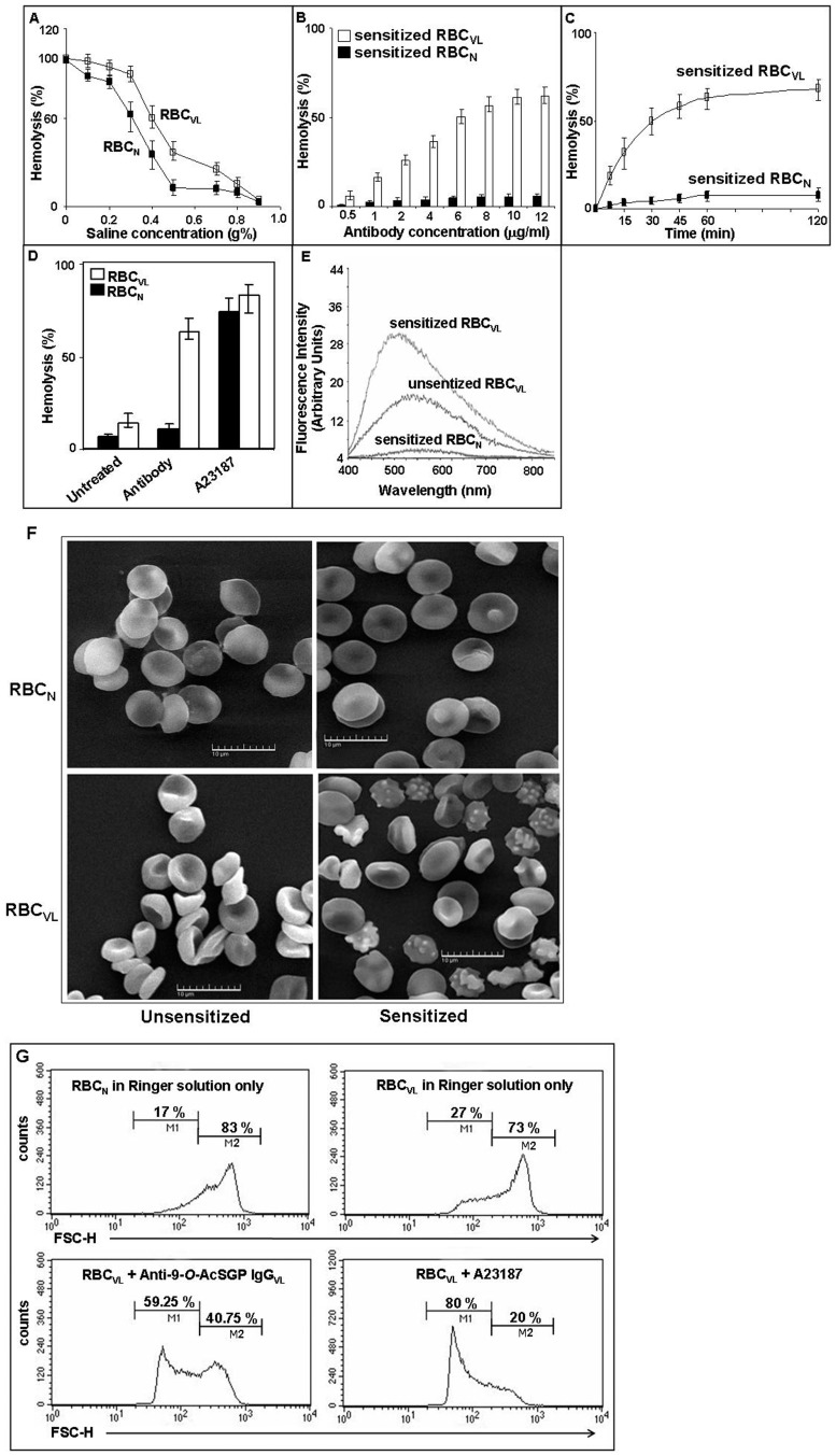
Figure 1. Altered membrane properties and reduced stability of RBCVL after sensitization with anti-9-O-AcSGP IgGVL antibodies.
A. The osmotic fragility of RBCVL (open square) compared to RBCN (filled square) was measured by degree of hemolysis with increasing osmotic stress by incubation in NaCl (0–0.9%) determined by spectrophotometry. Data are represented as % hemolysis, are means of triplicates ± SD and representative of three independent experiments. B. Sensitization of RBCVL for hemolysis with anti-9-O-AcSGP IgGVL antibodies. RBCVL (open bars) and RBCN (filled bars) were incubated for 30 min at 37°C with increasing concentration (0.5 µg/ml–12.0 µg/ml) of anti-9-O-AcSGP IgGVL and anti-9-O-AcSGP IgGNHS, respectively. The extent of hemolysis was then determined by spectrophotometry. The Spectrophotometric readings of RBCVL or RBCN in buffer under similar condition were taken as base values and subtracted from the experimental values. Data are expressed as means ± SD of three independent experiments. C. Kinetics of the hemolysis of RBCVL and RBCN after sensitization with anti-9-O-AcSGP IgGVL antibodies. RBCVL (open squares) and RBCN (filled squares) were incubated with equal (6.0 µg/ml) amounts of anti-9-O-AcSGP IgGVL and anti-9-O-AcSGP IgGNHS, respectively, for different time points (0–120 min). Base value was routinely subtracted from each experimental value as in Fig. 1B. Data are means ± SD of three independent experiments. D. Hemolysis of RBCN (filled bars) and RBCVL (open bars) after sensitization of RBCVL with anti-9-O-AcSGP IgGNHS or anti-9-O-AcSGP IgGVL (6.0 µg/ml), respectively, or incubation with the calcium ionophore A23187 (1 µM) in Ringer solution for 30 min. Data are means ± SD of three independent experiments. E. Enhanced membrane hydrophobicity of RBCVL after sensitization with anti-9-O-AcSGP IgGVL. The hydrophobicity was determined spectrofluorimetrically using ANS as indicator. RBCVL were incubated with anti-9-O-AcSGP IgGVL antibodies or PBS only for 30 min at 37°C. As controls, RBCN were incubated with anti-9-O-AcSGP IgGNHS. The cells were washed with PBS, loaded with ANS and incubated for 1 h at 37°C. The fluorescence emission spectra were recorded from 400 to 850 nm with excitation at 365 nm. The graphs are representatives of the results obtained from three independent experiments. F. Morphological changes of RBCVL after sensitization with anti-9-O-AcSGP IgGVL antibodies. RBCN and RBCVL were not sensitized or sensitized with anti-9-O-ACSGP IgGNHS or anti-9-O-AcSGP IgGVL antibodies, respectively, (6.0 µg/ml) for 30 min at 37°C, and then processed for SEM as described in Materials and Methods. Representative images from three independent experiments are shown. G. Decrease of RBCVL cell size after sensitization detected by low-angle scattering light flow cytometry (forward scatter, FCS). FCS was measured of RBCN (upper left panel) and of RBCVL without sensitization (upper right panel) or with sensitization with anti-9-O-AcSGP IgGVL (2.5 µg/ml) (lower left panel) or treatment with A23187 (1.0 µM) (lower right panel). Figure 1F is excluded from this article's CC-BY license. See the accompanying retraction notice for more information.

