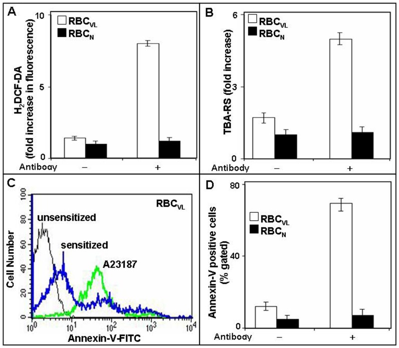
Figure 2. Enhanced ROS, lipid peroxidation and externalization of phosphatidyl serine (PS) in RBCVL after sensitization with anti-9-O-AcSGP IgGVL.
A. Induction of ROS in sensitized RBCVL. Prior to sensitization with anti-9-O-AcSGP IgGVL or anti-9-O-AcSGP IgGNHS RBCVL and RBCN were incubated with the hydroperoxide indicator H2DCF-DA in PBS for 30 min at 37°C. Then the antibodies were added, the cell incubated as before and ROS generation determined by fluorimetry. As controls, ROS generation was determined after prior treatment of the RBC with N-acetyl cysteine, a quencher of hydroperoxides. The results are expressed as means ± S.D. (n = 5) of fold increases in comparison to the fluorescence levels detected with untreated erythrocytes. B. Increased lipid peroxidation in sensitized RBCVL. Lipid peroxidation of erythrocyte membrane was detected and quantified with thiobarbituric acid (TBA) and the TBA-reactive species (TBA-RS) measured with a spectrophotometer at 532 nm. The results are mean ± S.D of fold increase in comparison with unsensitized RBC of five independent measurments. C–D. Enhanced externalization of phosphatidylserine on sensitized RBCVL. Sensitized or unsensitized erythrocytes were incubated in annexin-V binding buffer with FITC-annexin-V and analyzed by flow cytometry. A23187-treated RBC in the presence of Ca2+ served as positive control. The results are shown as representative histogram of three independent experiments (C) and as comparative flow-cytometric analysis (D).

