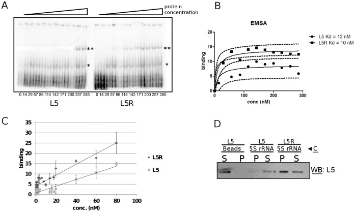
Figure 7. A mutation in the C-terminal domain of T. brucei L5 restoring a consensus arginine increases affinity for 5S rRNA.
Panel A: EMSA using radiolabeled 5S rRNA and recombinant T. brucei wild-type (left) and arginine mutant (right) proteins. The asterisk indicates the monomer:RNA complex and the double asterisk indicates the dimer:RNA complex. The numbers below indicate concentration of protein in nM. Panel B: Quantification of the data in Panel A. The dashed lines indicate 90% confidence intervals for the fitted curves. Panel C: Filter binding assay of reactions with recombinant L5 or L5R and radiolabeled 5S rRNA. The range of concentrations was chosen to represent binding to monomer L5. Panel D: RNA capture of recombinant L5 and L5R bound to biotinylated 5S rRNA. C: Capture, P: precipitate, S: supernatant.

