
Figure 1.
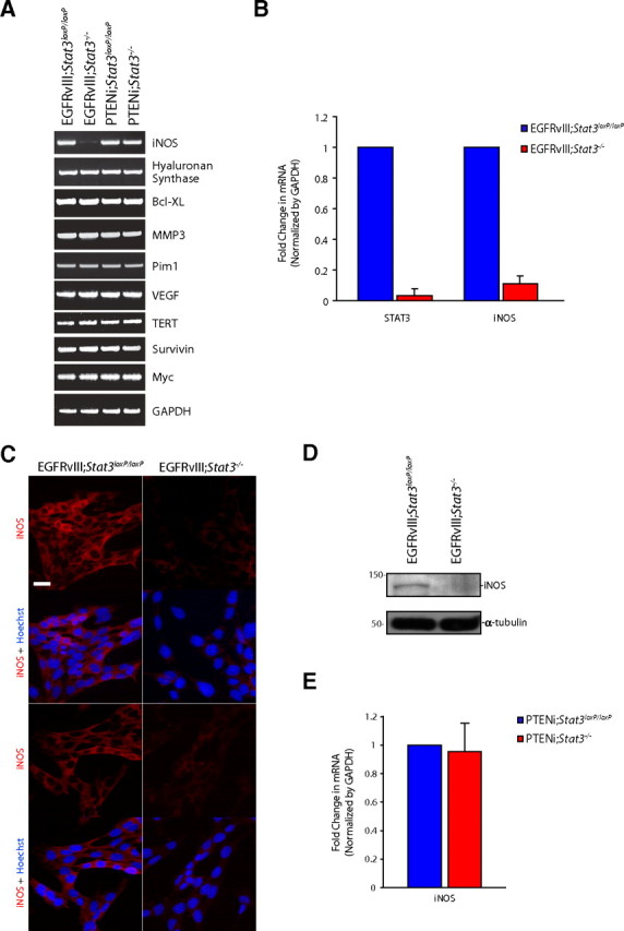
STAT3 regulates iNOS expression in EGFRvIII-expressing astrocytes. A, RNA was isolated from mouse EGFRvIII;Stat3loxP/loxP, EGFRvIII;Stat3−/−, PTENi;Stat3loxP/loxP, and PTENi;Stat3−/− astrocytes and subjected to RT-PCR with primers specific for the indicated genes. GAPDH served as control. iNOS mRNA levels were specifically reduced in EGFRvIII;Stat3−/− astrocytes compared with EGFRvIII;Stat3loxP/loxP astrocytes. B, RNA isolated from mouse EGFRvIII;Stat3loxP/loxP and EGFRvIII;Stat3−/− astrocytes was subjected to quantitative RT-PCR analyses using primers specific for STAT3 and iNOS. mRNA levels were normalized to GAPDH. STAT3 and iNOS mRNA levels were significantly reduced in EGFRvIII;Stat3−/− astrocytes compared with EGFRvIII;Stat3loxP/loxP astrocytes (ANOVA, p < 0.0005, n = 3). C, EGFRvIII;Stat3loxP/loxP and EGFRvIII;Stat3−/− astrocytes were subjected to immunocytochemistry using the rabbit iNOS antibody. Representative images are shown. The expression of iNOS was substantially reduced in EGFRvIII;Stat3−/− astrocytes compared with EGFRvIII;Stat3loxP/loxP astrocytes. Scale bar, 20 μm. D, Lysates of EGFRvIII;Stat3loxP/loxP and EGFRvIII;Stat3−/− astrocytes were immunoblotted with the iNOS or α-tubulin antibody. The levels of iNOS protein were substantially reduced in EGFRvIII;Stat3−/− astrocytes compared with EGFRvIII;Stat3loxP/loxP astrocytes. E, RNA isolated from mouse PTENi;Stat3loxP/loxP and PTENi;Stat3−/− astrocytes was subjected to quantitative RT-PCR analyses using primers specific for iNOS. mRNA levels were normalized to GAPDH. iNOS mRNA levels were not significantly different between PTENi;Stat3loxP/loxP astrocytes compared with PTENi;Stat3−/− astrocytes.