
Figure 2.
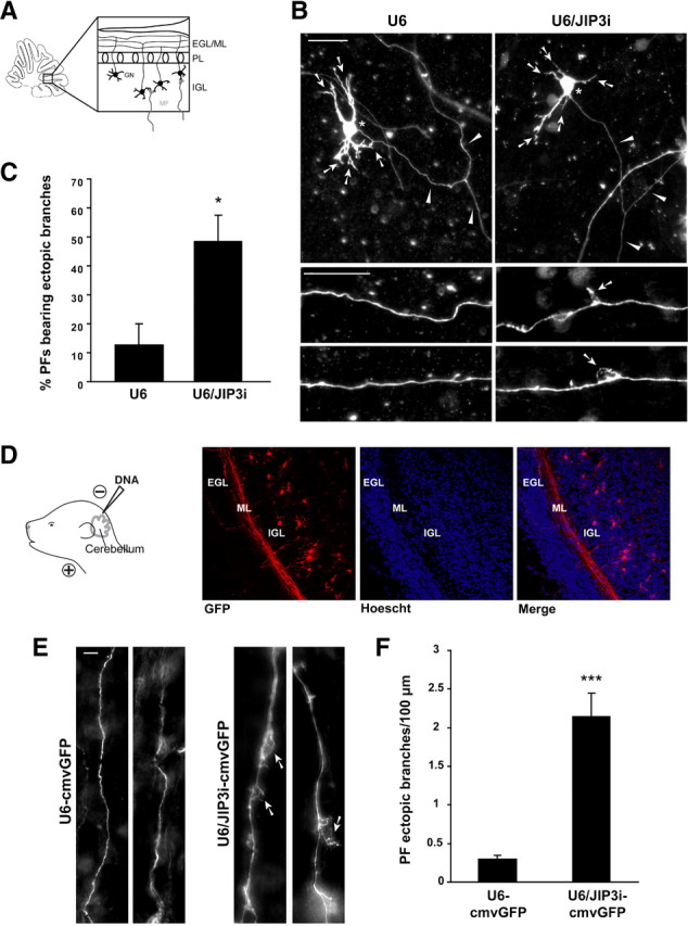
JIP3 suppresses axon branching in the cerebellar cortex in vivo. A, Schematic of cerebellar slice depicting organization of the cerebellar cortex, including the external granule layer/molecular layer (EGL/ML), the Purkinje cell layer (PL), and the internal granule layer (IGL). B, P9 rat cerebellar slices were transfected at DIV4 with the U6/JIP3i or control U6 RNAi plasmid together with the GFP expression plasmid, fixed 4 d later at DIV8, and subjected to immunohistochemistry using the GFP antibody. Top panels show examples of transfected granule neurons in each condition. Arrows indicate dendrites, asterisks the soma, and arrowheads the parallel fiber axon. Bottom panels show high-magnification images of distal axons in each condition. Arrows in bottom panels point to ectopic branches observed upon JIP3 knockdown. Scale bars, 25 μm. C, Quantification of slices treated as in B. JIP3 knockdown significantly increased the percentage of parallel fibers bearing one or more ectopic branches (t test, p < 0.05; n = 3). D, Left, Schematic of in vivo electroporation paradigm. Briefly, P3 rat pups were injected with DNA at the outer layer of the cerebellum and electroporated. Five days after electroporation, pups were killed and coronal cerebellar sections were subjected to immunohistochemistry using the GFP antibody. Right, Panels show a control section, with the EGL, ML, and IGL identified by Hoechst staining of nuclei. Overlay of GFP staining and Hoechst reveals that the majority of electroporated neurons reside in the IGL and extend axonal processes through the ML. E, High-magnification views of individual GFP-positive parallel fibers in the ML of U6–cmvGFP- or U6/JIP3i–cmvGFP-electroporated cerebella. Arrows indicate ectopic branches from the primary axonal fiber. Scale bar, 5 μm. F, Quantification of parallel fiber ectopic branches in the cerebellar cortex in control and JIP3 knockdown animals. The number of ectopic branches in parallel fibers was significantly increased in JIP3 knockdown animals compared with control animals (t test, p < 0.001). Measurements were collected from a total of 11 animals.