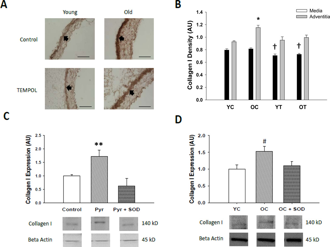
Figure 3. Collagen I protein expression.
(A) Representative immunohistochemistry images of collagen I in thoracic aortic sections from young and old control (YC and OC) and TEMPOL (YT and OT) supplemented mice. Arrows demarcate the medial-adventitial border; bar = 100 µm. (B) Collagen I protein in thoracic aortic sections of young and old control (YC and OC) and TEMPOL supplemented (YT and OT) mice. (C) Effects of pyrogallol (Pyr, 10 µM), polyethylene glycol conjugated superoxide dismutase (PEG-SOD, 20 U/mL) and the combination on collagen I adventitial fibroblasts from aorta of young rats. (D) Collagen I expression in adventitial fibroblasts from young and old control rats (YC and OC) and effects of PEG-SOD (20 U/mL) in fibroblasts from old rats (OC + SOD). Values are mean ± SEM. (n = 5 – 7 per group) * p < 0.05 for Adventitia OC vs. Adventitia YC, Adventitia YT and Adventitia OT. † p < 0.05 for Media YT vs. Media YC and Media OC, Media OT vs. Media YC and Media OC. ** p < 0.05 for Pyr vs. control, Pry + SOD. # p < 0.05 for OC vs. YC and OC + SOD.

